Jung Su-Kyung, Lee Edward H, Mishra Kavita K, Daftari Inder K, Park Susanna S
Department of Ophthalmology and Vision Science, University of California Davis Eye Center, Sacramento, California.
Department of Ophthalmology, Hospital, National Cancer Center, Gyeonggi-do, Korea.
Ophthalmol Sci. 2024 Dec 12;5(3):100674. doi: 10.1016/j.xops.2024.100674. eCollection 2025 May-Jun.
To evaluate the macular and peripapillary retinal and choroidal flow changes in eyes with choroidal melanoma (CM) treated with proton beam radiation therapy (PBRT) using OCT angiography (OCTA).
A prospective, cross-sectional, single-center study.
All patients seen at the study center between 2019 and 2024 who received PBRT for CM in 1 eye ≥1 year before enrollment with best-corrected visual acuity (BCVA) >20/200, unremarkable contralateral eye, and agreed to participate.
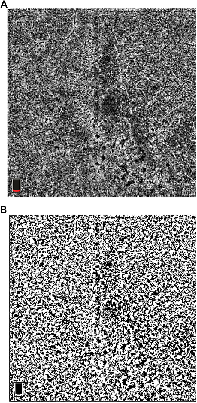
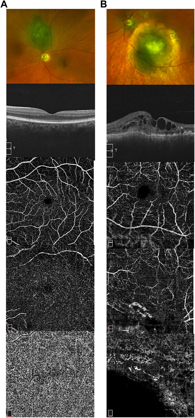
After a comprehensive eye examination, including BCVA, Optovue AngioVue was used to obtain the 4.5-mm optic disc and 6.0-mm macular OCT/OCT angiography (OCTA) images of both eyes. All vascular density (VD) measurements were obtained automatically using the OCTA software, except choriocapillaris VD, which was quantitated using ImageJ. The Wilcoxon signed-rank test was used to analyze differences in OCT/OCTA parameters between the treated and the contralateral eyes. Spearman's ρ was used to identify OCTA parameters associated with BCVA or radiation dose. A value of <0.05 was considered statistically significant.
Foveal avascular zone (FAZ) area and perimeter, choriocapillaris and retinal (superficial and deep) capillary VD in the macula and radial peripapillary capillary (RPC) VD on OCTA; macular and retinal nerve fiber layer thickness on OCT, tumor location, laterality and size at baseline, BCVA of both eyes, PBRT dose, and duration of follow-up at enrollment.
Among 24 participants, OCT/OCTA parameters were significantly different in the treated eyes when compared with the contralateral eyes, including increased FAZ area and perimeter, decreased peripapillary retinal nerve fiber layer thickness and RPC VD, and decreased macular choriocapillaris VD and parafoveal and perifoveal superficial retinal plexus VD ( < 0.05). Best-corrected visual acuity in the treated eyes correlated significantly with FAZ area and perimeter, parafoveal and perifoveal deep retinal plexus VD, and radiation dose to fovea but not radiation dose to the optic disc.
Although PBRT can affect both retinal and choroidal vascular flow in the macular and peripapillary region in eyes with CM, BCVA after PBRT seems to correlate best with the retinal vascular flow changes in the macula on OCTA and radiation dose to the fovea.
Proprietary or commercial disclosure may be found in the Footnotes and Disclosures at the end of this article.
使用光学相干断层扫描血管造影(OCTA)评估接受质子束放射治疗(PBRT)的脉络膜黑色素瘤(CM)患者眼内黄斑区、视乳头周围视网膜和脉络膜的血流变化。
一项前瞻性、横断面、单中心研究。
2019年至2024年期间在研究中心就诊的所有患者,这些患者在入组前至少1年有1只眼因CM接受了PBRT,最佳矫正视力(BCVA)>20/200,对侧眼无异常,且同意参与研究。
在进行包括BCVA在内的全面眼部检查后,使用Optovue AngioVue获取双眼4.5毫米视盘和6.0毫米黄斑区的OCT/OCT血管造影(OCTA)图像。除脉络膜毛细血管血管密度(VD)使用ImageJ定量外,所有血管密度测量均使用OCTA软件自动获取。采用Wilcoxon符号秩检验分析治疗眼与对侧眼之间OCT/OCTA参数的差异。使用Spearman's ρ来确定与BCVA或放射剂量相关的OCTA参数。P < 0.05被认为具有统计学意义。
OCTA上黄斑中心凹无血管区(FAZ)面积和周长、黄斑区脉络膜毛细血管和视网膜(浅层和深层)毛细血管VD以及视乳头周围放射状毛细血管(RPC)VD;OCT上黄斑和视网膜神经纤维层厚度、肿瘤位置、基线时的患侧性和大小、双眼BCVA、PBRT剂量以及入组时的随访时间。
24名参与者中,治疗眼的OCT/OCTA参数与对侧眼相比有显著差异,包括FAZ面积和周长增加、视乳头周围视网膜神经纤维层厚度和RPC VD降低、黄斑区脉络膜毛细血管VD以及黄斑旁和黄斑周围浅层视网膜丛VD降低(P < 0.05)。治疗眼的最佳矫正视力与FAZ面积和周长、黄斑旁和黄斑周围深层视网膜丛VD以及黄斑中心凹的放射剂量显著相关,但与视盘的放射剂量无关。
虽然PBRT可影响CM患者眼内黄斑区和视乳头周围区域的视网膜和脉络膜血流,但PBRT后的BCVA似乎与OCTA上黄斑区视网膜血流变化以及黄斑中心凹的放射剂量相关性最佳。
本文末尾的脚注和披露中可能会有专有或商业披露信息。