Talarico M, Barbato S, Cattabriga A, Sacchetti I, Manzato E, Restuccia R, Masci S, Bigi F, Puppi M, Iezza M, Rizzello I, Mancuso K, Pantani L, Tacchetti P, Nanni C, Cavo M, Zamagni E
IRCCS Azienda Ospedaliero-Universitaria di Bologna, Istituto di Ematologia "Seràgnoli", Bologna, Italy.
Dipartimento di Scienze Mediche e Chirurgiche, Università di Bologna, Bologna, Italy.
J Bone Oncol. 2025 Feb 28;51:100669. doi: 10.1016/j.jbo.2025.100669. eCollection 2025 Apr.
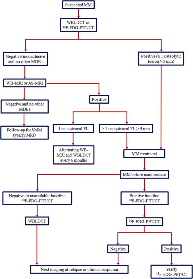
The International Myeloma Working Group (IMWG) defines myeloma related bone disease (MBD) as a diagnostic criterion for symptomatic multiple myeloma (MM) as the presence of osteolytic lesions ≥ 5 mm or more than one focal lesion (FL) ≥ 5 mm by magnetic resonance imaging (MRI). Whole-body low-dose CT (WBLDCT) is recommended as the first-choice imaging technique for the diagnosis of MBD with F-fluorodeoxyglucose-positron emission tomography/CT (F-FDG-PET/CT) being considered a possible alternative at staging, whereas use of MRI studies is recommended in cases without myeloma-defining events (MDEs) in order to exclude the presence of FLs. Furthermore, use of F-FDG-PET/CT is recommended in response assessment, to be integrated with hematologic response and bone marrow minimal residual disease (MRD).
In this paper, we review novel functional imaging techniques in MM, particularly focusing on their advantages, limits, applications and comparisons with F-FDG-PET/CT or other standardized imaging techniques.
Combining both morphological and functional imaging, F-FDG-PET/CT is currently considered a standard imaging technique in MM for staging (despite false positive or negative results) and response assessment. The introduction of novel functional imaging techniques, as whole-body diffusion-weighted magnetic resonance imaging (WB-DWI-MRI), or novel PET tracers might be useful in overcoming these limits. Future studies will give more information on the complementarity of these imaging techniques or whether one of them might become a new gold standard in MM.
国际骨髓瘤工作组(IMWG)将骨髓瘤相关骨病(MBD)定义为有症状的多发性骨髓瘤(MM)的诊断标准,即通过磁共振成像(MRI)发现溶骨性病变≥5mm或超过一个局灶性病变(FL)≥5mm。全身低剂量CT(WBLDCT)被推荐作为诊断MBD的首选成像技术,F-氟脱氧葡萄糖正电子发射断层扫描/CT(F-FDG-PET/CT)在分期时被认为是一种可能的替代方法,而对于无骨髓瘤定义事件(MDE)的病例,建议使用MRI检查以排除FL的存在。此外,在疗效评估中推荐使用F-FDG-PET/CT,并将其与血液学缓解和骨髓微小残留病(MRD)相结合。
在本文中,我们回顾了MM中的新型功能成像技术,特别关注它们的优势、局限性、应用以及与F-FDG-PET/CT或其他标准化成像技术的比较。
结合形态学和功能成像,F-FDG-PET/CT目前被认为是MM分期(尽管存在假阳性或假阴性结果)和疗效评估的标准成像技术。引入新型功能成像技术,如全身扩散加权磁共振成像(WB-DWI-MRI)或新型PET示踪剂,可能有助于克服这些局限性。未来的研究将提供更多关于这些成像技术互补性的信息,或者其中一种是否可能成为MM的新金标准。