Blandinières Adeline, Rossi Elisa, Gendron Nicolas, Rancic Jeanne, Rosa Mickael, Dupont Annabelle, Idelcadi Salim, Philippe Aurélien, Poitier Bastien, Bièche Ivan, Vacher Sophie, Cholley Bernard, Gaussem Pascale, Susen Sophie, Smadja David M
Université Paris Cité, Innovative Therapies in Haemostasis, INSERM, Paris, France.
AP-HP, European Georges Pompidou Hospital, Hematology Department, Paris, France.
J Cell Mol Med. 2025 Apr;29(7):e70511. doi: 10.1111/jcmm.70511.
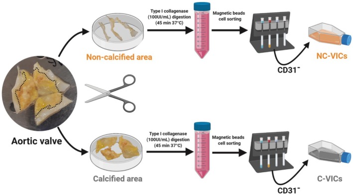
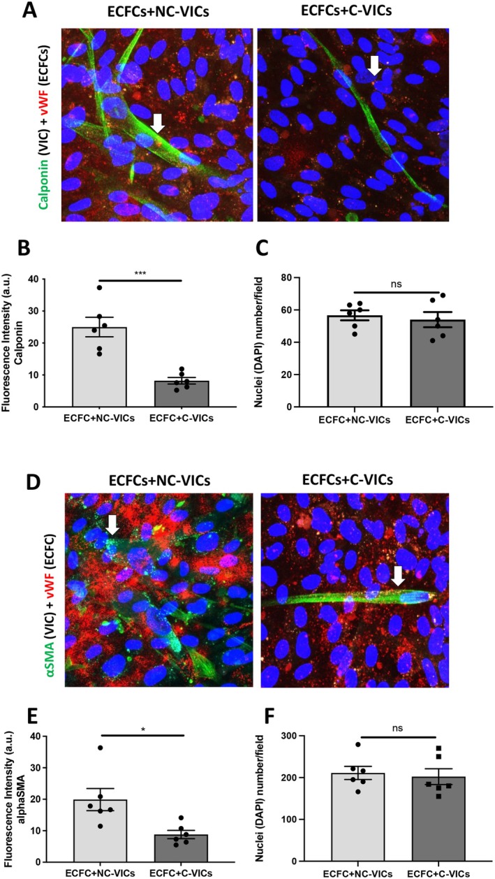
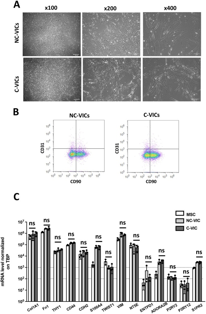
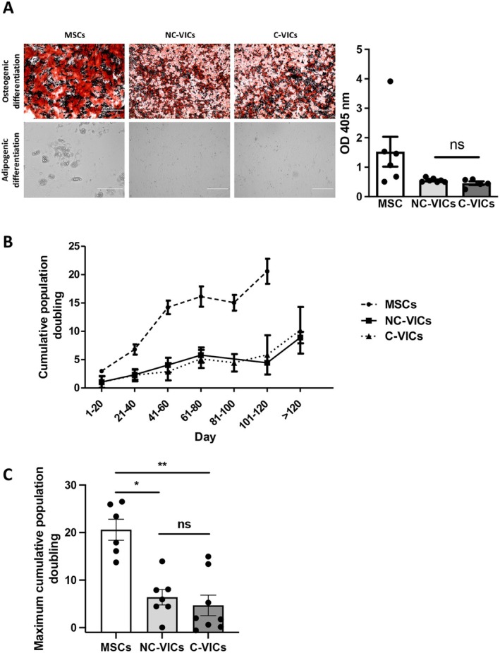
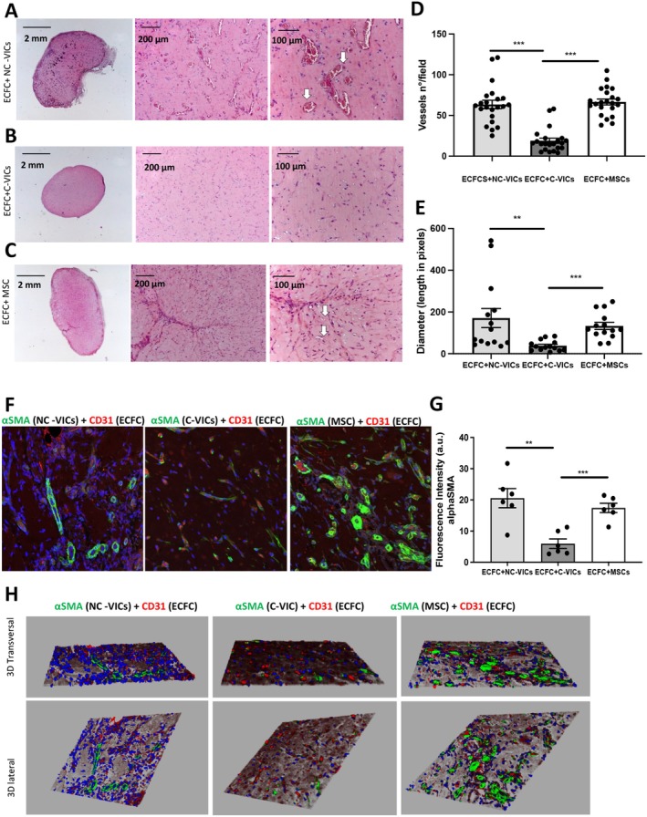
Valve interstitial cells (VICs) play a critical role in aortic valve calcification and angiogenic processes associated with calcific aortic valve stenosis (CAVS). Within the same valve, VICs from differently calcified regions can exhibit diverse phenotypic and functional properties. We hypothesised that VICs isolated from noncalcified (NC-VICs) and calcified (C-VICs) areas of human aortic valves possess distinct angiogenic characteristics. In this study, we isolated C-VICs and NC-VICs from 23 valves obtained after aortic valve replacement due to CAVS. Both VIC types exhibited similar phenotypes in culture, characterised by morphology, expression of mesenchymal/fibroblastic markers, proliferation and osteogenic differentiation. No significant differences were observed in the secretion of angiogenic factors, including VEGF-A, Ang-1, Ang-2, PlGF, bFGF between NC-VICs and C-VICs. However, when co-injected with endothelial colony-forming cells (ECFCs) into Matrigel implants in vivo in mice, implants containing NC-VICs showed significantly higher microvessel density compared to those with C-VICs (p < 0.001). Additionally, NC-VICs co-cultured with ECFCs expressed significantly higher levels of the perivascular markers αSMA and calponin compared to C-VICs (p < 0.001 and p < 0.05, respectively). In conclusion, our study reveals the heterogeneity in VIC plasticity within the aortic valve during CAVS. The diminished capacity of VICs from calcified areas to differentiate into perivascular cells suggests a loss of function as valve disease progresses. Furthermore, the ability of VICs to undergo perivascular differentiation may provide insights into valve homeostasis, angiogenesis and the exacerbation of calcification.
瓣膜间质细胞(VICs)在主动脉瓣钙化以及与钙化性主动脉瓣狭窄(CAVS)相关的血管生成过程中起着关键作用。在同一瓣膜内,来自不同钙化区域的VICs可表现出不同的表型和功能特性。我们推测,从人主动脉瓣非钙化(NC-VICs)和钙化(C-VICs)区域分离出的VICs具有不同的血管生成特征。在本研究中,我们从因CAVS行主动脉瓣置换术后获得的23个瓣膜中分离出C-VICs和NC-VICs。两种VIC类型在培养中表现出相似的表型,其特征为形态、间充质/成纤维细胞标志物的表达、增殖和成骨分化。在NC-VICs和C-VICs之间,包括VEGF-A、Ang-1、Ang-2、PlGF、bFGF在内的血管生成因子分泌未观察到显著差异。然而,当与内皮集落形成细胞(ECFCs)共同注射到小鼠体内的基质胶植入物中时,含有NC-VICs的植入物与含有C-VICs的植入物相比,微血管密度显著更高(p < 0.001)。此外,与C-VICs相比,与ECFCs共培养的NC-VICs表达的血管周标志物αSMA和钙调蛋白水平显著更高(分别为p < 0.001和p < 0.05)。总之,我们的研究揭示了CAVS期间主动脉瓣内VIC可塑性的异质性。钙化区域的VICs分化为血管周细胞的能力减弱表明,随着瓣膜疾病进展功能丧失。此外,VICs进行血管周分化的能力可能为瓣膜稳态、血管生成和钙化加剧提供见解。