Biomedical Engineering Department, Worcester Polytechnic Institute, Worcester, MA 01609, USA.
Cells. 2023 Dec 25;13(1):45. doi: 10.3390/cells13010045.
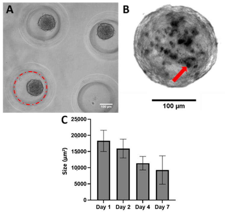
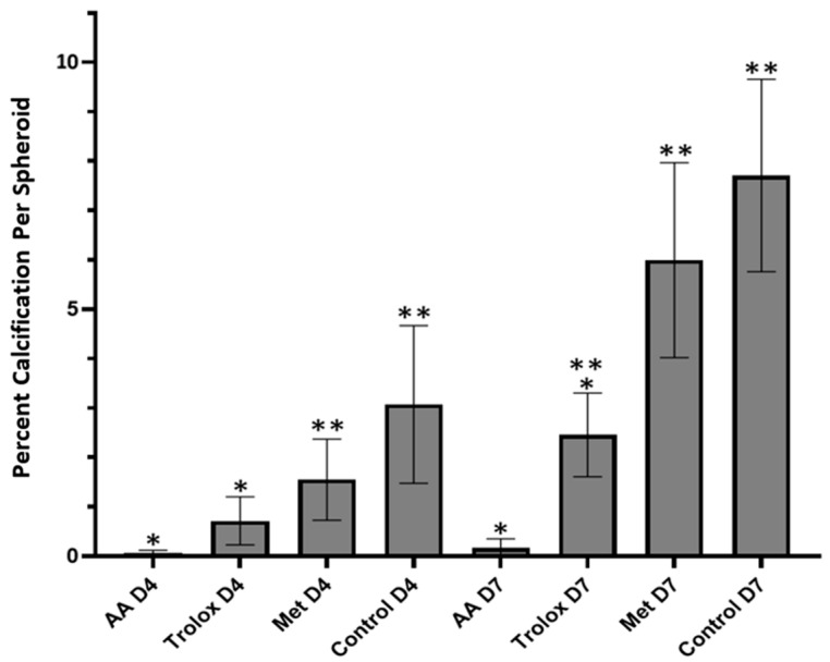
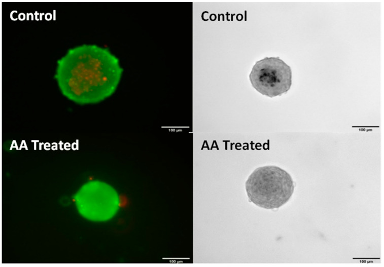
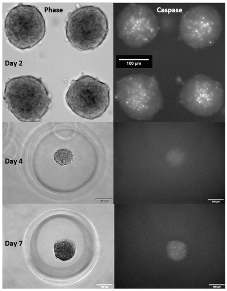
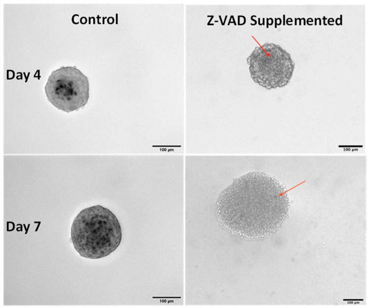
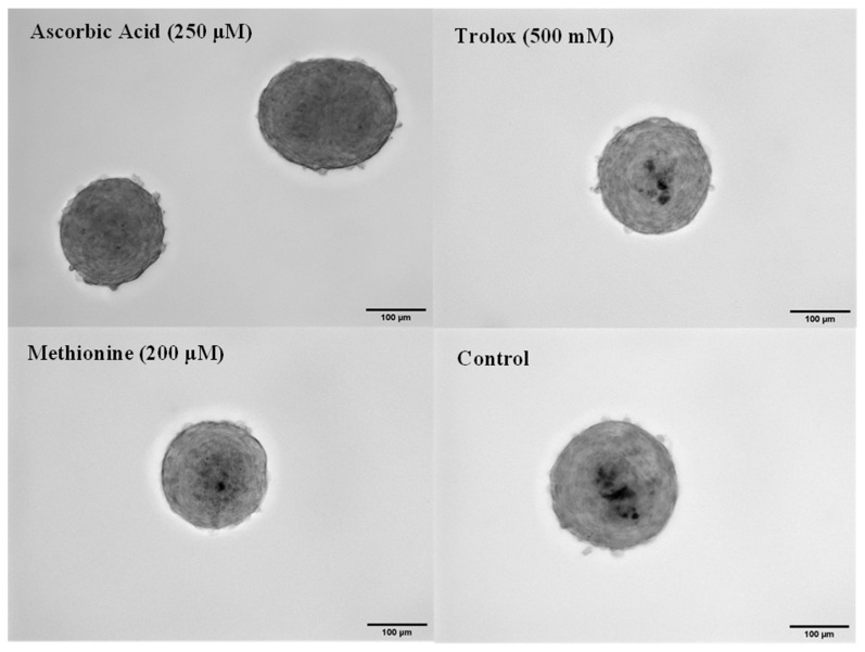
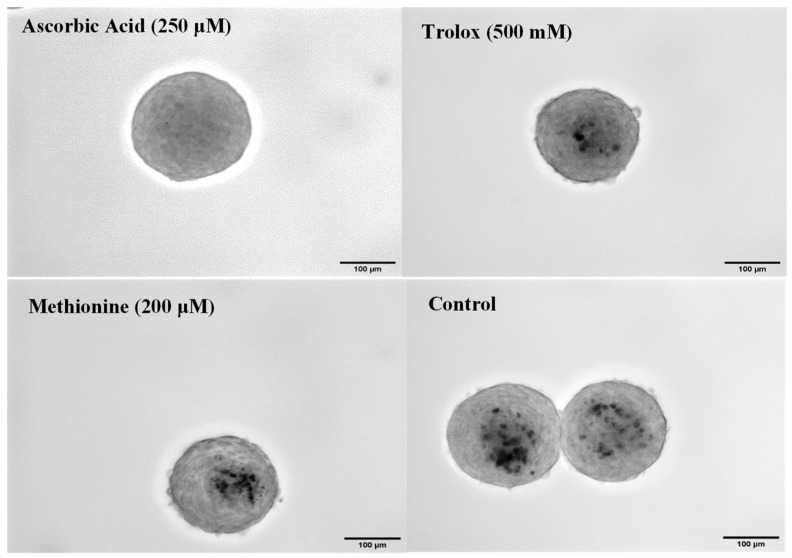
Calcific aortic valve disease (CAVD) is the most common heart valve disease among aging populations. There are two reported pathways of CAVD: osteogenic and dystrophic, the latter being more prevalent. Current two-dimensional (2D) in vitro CAVD models have shed light on the disease but lack three-dimensional (3D) cell-ECM interactions, and current 3D models require osteogenic media to induce calcification. The goal of this work is to develop a 3D dystrophic calcification model. We hypothesize that, as with 2D cell-based CAVD models, programmed cell death (apoptosis) is integral to calcification. We model the cell aggregation observed in CAVD by creating porcine valvular interstitial cell spheroids in agarose microwells. Upon culture in complete growth media (DMEM with serum), calcium nodules form in the spheroids within a few days. Inhibiting apoptosis with Z-VAD significantly reduced calcification, indicating that the calcification observed in this model is dystrophic rather than osteogenic. To determine the relative roles of oxidative stress and extracellular matrix (ECM) production in the induction of apoptosis and subsequent calcification, the media was supplemented with antioxidants with differing effects on ECM formation (ascorbic acid (AA), Trolox, or Methionine). All three antioxidants significantly reduced calcification as measured by Von Kossa staining, with the percentages of calcification per area of AA, Trolox, Methionine, and the non-antioxidant-treated control on day 7 equaling 0.17%, 2.5%, 6.0%, and 7.7%, respectively. As ZVAD and AA almost entirely inhibit calcification, apoptosis does not appear to be caused by a lack of diffusion of oxygen and metabolites within the small spheroids. Further, the observation that AA treatment reduces calcification significantly more than the other antioxidants indicates that the ECM stimulatory effect of AA plays a role inhibiting apoptosis and calcification in the spheroids. We conclude that, in this 3D in vitro model, both oxidative stress and ECM production play crucial roles in dystrophic calcification and may be viable therapeutic targets for preventing CAVD.
钙化性主动脉瓣疾病(CAVD)是老年人群中最常见的心脏瓣膜病。CAVD 有两种报道的途径:成骨性和营养不良性,后者更为普遍。目前的二维(2D)体外 CAVD 模型已经揭示了该疾病,但缺乏三维(3D)细胞-细胞外基质相互作用,并且目前的 3D 模型需要成骨介质来诱导钙化。这项工作的目标是开发一种 3D 营养不良性钙化模型。我们假设,与 2D 基于细胞的 CAVD 模型一样,程序性细胞死亡(细胞凋亡)是钙化的组成部分。我们通过在琼脂糖微井中创建猪瓣膜间质细胞球体来模拟 CAVD 中观察到的细胞聚集。在完全生长培养基(含血清的 DMEM)中培养几天后,球体中形成钙结节。用 Z-VAD 抑制细胞凋亡显著减少了钙化,表明该模型中观察到的钙化是营养不良性的而不是成骨性的。为了确定氧化应激和细胞外基质(ECM)产生在诱导细胞凋亡和随后的钙化中的相对作用,用对 ECM 形成有不同影响的抗氧化剂(抗坏血酸(AA)、Trolox 或蛋氨酸)补充培养基。所有三种抗氧化剂均通过 Von Kossa 染色显著减少钙化,第 7 天 AA、Trolox、蛋氨酸和非抗氧化剂处理对照的面积百分比的钙化分别为 0.17%、2.5%、6.0%和 7.7%。由于 ZVAD 和 AA 几乎完全抑制钙化,因此细胞凋亡似乎不是由于小球体内氧气和代谢物扩散不足引起的。此外,AA 处理可显著降低钙化的观察结果表明,AA 的 ECM 刺激作用在抑制球体中的细胞凋亡和钙化中起作用。我们得出结论,在这个 3D 体外模型中,氧化应激和 ECM 产生都在营养不良性钙化中起关键作用,并且可能是预防 CAVD 的可行治疗靶标。