Xu Fangfang, Zhang Ying, Ma Qianqing, Hu Lili, Li Yu, Gao Chuanfen, Guo Peipei, Yang Xianyue, Zhou Yi, Zhang Jie, Wang Heng, Zhang Chaoxue
Department of Ultrasound, The First Affiliated Hospital of Anhui Medical University, No. 218 Jixi Road, Shushan District, Hefei, Anhui, 230022, People's Republic of China.
Hefei Maternal and Child Health Hospital, Hefei, China.
BMC Pregnancy Childbirth. 2025 Apr 3;25(1):391. doi: 10.1186/s12884-025-07508-0.
To investigate the optimal periendometrial zone (PEZ) in ultrasound (US) images and assess the performance of ultrasound radiomics in predicting the outcome of frozen embryo transfer (FET).
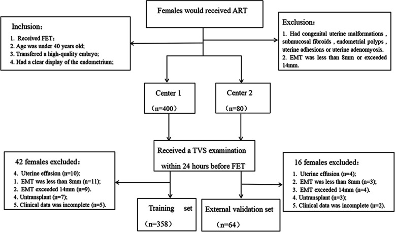
This prospective study had 422 female participants (training set: n = 358, external validation set: n = 64). We delineated the region of interest (ROI) of the endometrium (EN) from ultrasound images of the median sagittal surface of the uteri of patients. We determined the ROIs of PEZ on US images by automatically expanding 2.0, 4.0, 6.0, and 8.0 mm radii surrounding the EN. We determined the radiomics characteristics based on the ROIs of the endometrium and PEZ, then compared the different sizes of PEZ to determine the optimal PEZ. We constructed models of the EN and optimal PEZ using six machine learning algorithms. We developed a combined model using the radiomics characteristics of EN and the optimal PEZ. We evaluated the performance of the three models using the area under the curve (AUC).
The optimal PEZ was 4.0 mm with a maximum AUC of 0.715 (95% confidence interval (CI): 0.581 - 0.833) in the external validation set. The combined radiomics model (endometrium and PEZ) yielded the best predictive performance with AUC = 0.853 (95% CI: 0.811 - 0.890) for the training set and AUC = 0.809 (95% CI: 0.696 - 0.909) for the external validation set.
PEZ could be the optimal area for predicting clinical pregnancy after FET. An US-based radiomics model that combines EN and PEZ demonstrated strong potential in helping clinicians predict FET outcomes more accurately, thereby supporting informed decision-making before treatment.
研究超声(US)图像中最佳的子宫内膜周围区(PEZ),并评估超声影像组学在预测冻融胚胎移植(FET)结局方面的性能。
这项前瞻性研究纳入了422名女性参与者(训练集:n = 358,外部验证集:n = 64)。我们从患者子宫正中矢状面的超声图像中勾勒出子宫内膜(EN)的感兴趣区域(ROI)。通过在EN周围自动扩展半径为2.0、4.0、6.0和8.0 mm来确定US图像上PEZ的ROI。我们基于子宫内膜和PEZ的ROI确定影像组学特征,然后比较不同大小的PEZ以确定最佳PEZ。我们使用六种机器学习算法构建了EN和最佳PEZ的模型。我们利用EN和最佳PEZ的影像组学特征开发了一个联合模型。我们使用曲线下面积(AUC)评估这三个模型的性能。
在外部验证集中,最佳PEZ为4.0 mm,最大AUC为0.715(95%置信区间(CI):0.581 - 0.833)。联合影像组学模型(子宫内膜和PEZ)产生了最佳预测性能,训练集的AUC = 0.853(95% CI:0.811 - 0.890),外部验证集的AUC = 0.809(95% CI:0.696 - 0.909)。
PEZ可能是预测FET后临床妊娠的最佳区域。基于超声的结合EN和PEZ的影像组学模型在帮助临床医生更准确地预测FET结局方面显示出强大潜力,从而支持治疗前的明智决策。