Dodo Cindy, Senna Plinio Mendes, Del Bel Cury Altair Antoninha, Meirelles Luiz
Department of General Dental Sciences, School of Dentistry, Marquette University, Milwaukee, Wisconsin, USA.
Department of Prosthodontics, School of Dentistry, Rio de Janeiro State University, Rio de Janeiro, Brazil.
Clin Implant Dent Relat Res. 2025 Apr;27(2):e70030. doi: 10.1111/cid.70030.
The long-term success of dental implants depends on the preservation of supporting tissues over time. Recent studies have highlighted the release of titanium particles as a potential etiology for the onset and progression of peri-implant diseases modulated by inflammatory biomarkers. This study provides a comprehensive analysis of surface changes associated with high insertion torque placement.
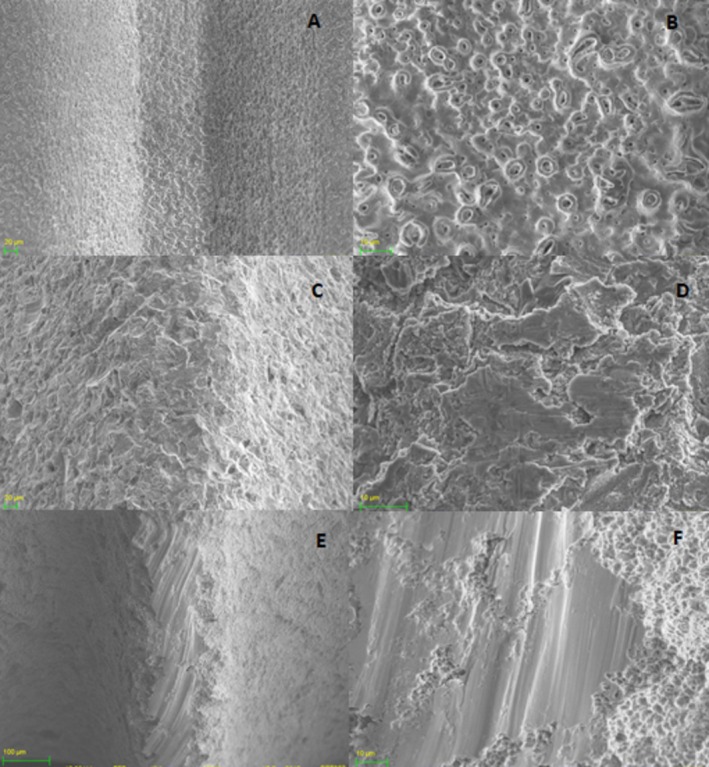
Three groups of cylindrical threaded dental implants, each representing different surface topographies produced by anodization or a combination of grit-blasting and acid-etching processes, were inserted into fresh cow rib bone blocks used to mimic human jaws. Individual bone blocks were fabricated with a dimension of 20 × 15 × 15 mm, randomly assigned to the three implant groups. Prior to dental implant placement, the bone blocks were divided in half to facilitate implant removal without introducing additional damage. The drilling protocol was modified, excluding the final drill recommended by the manufacturer to ensure higher insertion torque values during the procedure. Dental implants were removed from the bone blocks and processed for analysis. Surface roughness was characterized using interferometry on the same area before and after insertion. Scanning electron microscopy (SEM) with a back-scattered electron detector (BSD) was employed to identify the implant surface and loose particles at the bone block interface.
The high insertion torque protocol used in this study resulted in higher insertion torque values compared to manufacturers' protocol, but no difference was observed when comparing the three implant groups. Surface roughness characterization revealed that amplitude and hybrid roughness parameters for all three groups were lower after insertion. The surfaces exhibiting a predominance of peaks (Ssk [skewness] > 0) associated with higher structures (height parameters) showed greater damage at the crests of the threads, while no changes were observed in the valleys of the threads. SEM-BSD images revealed loose titanium particles at the bone blocks interface, predominantly at the crestal cortical bone level.
High insertion torque resulted in surface damage at the crests of threads, which subsequently led to the release of titanium particles primarily at the bone crest. The initial release of titanium particles during implant insertion at the bone-implant interface warrants further exploration as a potential cofactor for marginal bone loss.
牙种植体的长期成功取决于支持组织随时间的保存情况。最近的研究强调了钛颗粒的释放是由炎症生物标志物调节的种植体周围疾病发生和进展的潜在病因。本研究对与高植入扭矩放置相关的表面变化进行了全面分析。
三组圆柱形螺纹牙种植体,每组代表通过阳极氧化或喷砂和酸蚀工艺组合产生的不同表面形貌,被插入用于模拟人类颌骨的新鲜牛肋骨块中。单个骨块的尺寸为20×15×15毫米,随机分配到三个种植体组。在植入牙种植体之前,将骨块分成两半,以便于在不造成额外损伤的情况下取出种植体。修改了钻孔方案,排除了制造商推荐的最终钻头,以确保在手术过程中获得更高的植入扭矩值。将牙种植体从骨块中取出并进行分析处理。在植入前后的同一区域使用干涉测量法对表面粗糙度进行表征。使用带有背散射电子探测器(BSD)的扫描电子显微镜(SEM)来识别种植体表面和骨块界面处的松散颗粒。
与制造商的方案相比,本研究中使用的高植入扭矩方案导致更高的植入扭矩值,但在比较三个种植体组时未观察到差异。表面粗糙度表征显示,所有三组的幅度和混合粗糙度参数在植入后均较低。显示与较高结构(高度参数)相关的峰值占主导(偏度[Ssk]>0)的表面在螺纹顶部显示出更大的损伤,而在螺纹谷中未观察到变化。SEM-BSD图像显示在骨块界面处有松散的钛颗粒,主要在牙槽嵴皮质骨水平。
高植入扭矩导致螺纹顶部的表面损伤,随后主要在牙槽嵴处导致钛颗粒的释放。植入过程中在骨-种植体界面处钛颗粒的初始释放作为边缘骨丢失的潜在辅助因素值得进一步探索。