• 文献检索
  • 文档翻译
  • 深度研究
  • 学术资讯
  • Suppr Zotero 插件Zotero 插件
  • 邀请有礼
  • 套餐&价格
  • 历史记录
应用&插件
Suppr Zotero 插件Zotero 插件浏览器插件Mac 客户端Windows 客户端微信小程序
定价
高级版会员购买积分包购买API积分包
服务
文献检索文档翻译深度研究API 文档MCP 服务
关于我们
关于 Suppr公司介绍联系我们用户协议隐私条款
关注我们

Suppr 超能文献

核心技术专利:CN118964589B侵权必究
粤ICP备2023148730 号-1Suppr @ 2026

文献检索

告别复杂PubMed语法,用中文像聊天一样搜索,搜遍4000万医学文献。AI智能推荐,让科研检索更轻松。

立即免费搜索

文件翻译

保留排版,准确专业,支持PDF/Word/PPT等文件格式,支持 12+语言互译。

免费翻译文档

深度研究

AI帮你快速写综述,25分钟生成高质量综述,智能提取关键信息,辅助科研写作。

立即免费体验

钛植入物的缩放会引发炎症诱导的骨质溶解。

Scaling of titanium implants entrains inflammation-induced osteolysis.

机构信息

Department of Anatomy &Anthropology, Sackler Faculty of Medicine, Tel Aviv University, Israel.

Department of Prosthodontics, Goldschleger School of Dental Medicine, Sackler Faculty of Medicine, Tel Aviv University, Israel.

出版信息

Sci Rep. 2017 Jan 6;7:39612. doi: 10.1038/srep39612.

DOI:10.1038/srep39612
PMID:28059080
原文链接:https://pmc.ncbi.nlm.nih.gov/articles/PMC5216395/
Abstract

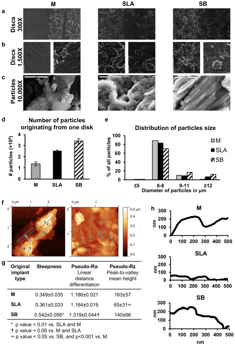
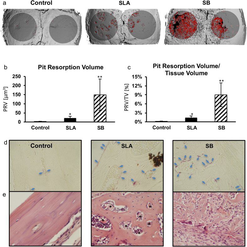
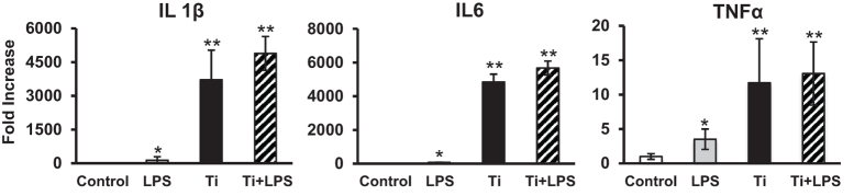
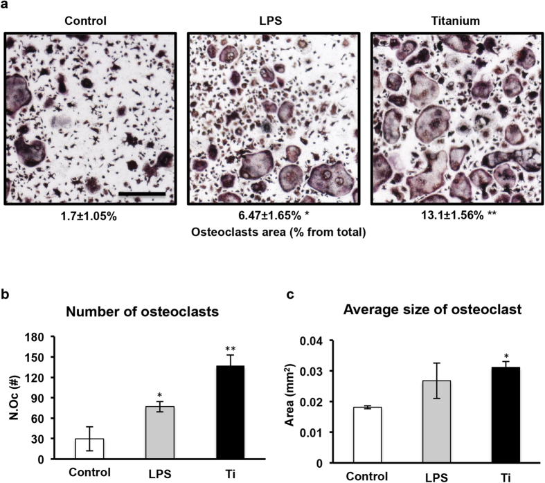
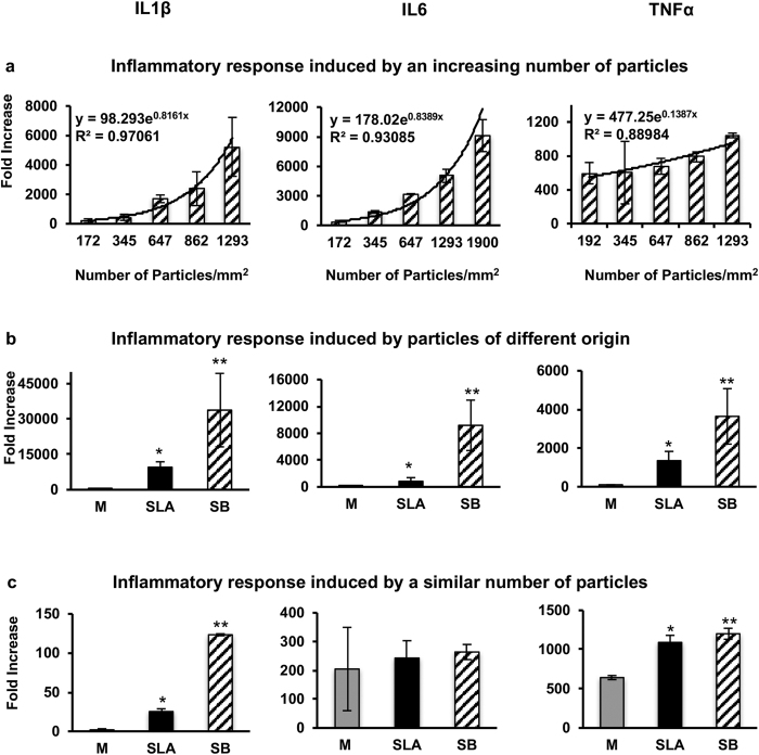
With millions of new dental and orthopedic implants inserted annually, periprosthetic osteolysis becomes a major concern. In dentistry, peri-implantitis management includes cleaning using ultrasonic scaling. We examined whether ultrasonic scaling releases titanium particles and induces inflammation and osteolysis. Titanium discs with machined, sandblasted/acid-etched and sandblasted surfaces were subjected to ultrasonic scaling and we physically and chemically characterized the released particles. These particles induced a severe inflammatory response in macrophages and stimulated osteoclastogenesis. The number of released particles and their chemical composition and nanotopography had a significant effect on the inflammatory response. Sandblasted surfaces released the highest number of particles with the greatest nanoroughness properties. Particles from sandblasted/acid-etched discs induced a milder inflammatory response than those from sandblasted discs but a stronger inflammatory response than those from machined discs. Titanium particles were then embedded in fibrin membranes placed on mouse calvariae for 5 weeks. Using micro-CT, we observed that particles from sandblasted discs induced more osteolysis than those from sandblasted/acid-etched discs. In summary, ultrasonic scaling of titanium implants releases particles in a surface type-dependent manner and may aggravate peri-implantitis. Future studies should assess whether surface roughening affects the extent of released wear particles and aseptic loosening of orthopedic implants.

摘要

每年有数百万个新的牙科和骨科植入物被植入,因此假体周围骨溶解成为一个主要关注点。在牙科领域,种植体周围炎的治疗包括使用超声洁治进行清洁。我们研究了超声洁治是否会释放钛颗粒,并引发炎症和骨溶解。对经过机械加工、喷砂酸蚀和喷砂处理的钛盘进行了超声洁治,并对释放的颗粒进行了物理和化学特性分析。这些颗粒在巨噬细胞中引发了严重的炎症反应,并刺激破骨细胞生成。释放颗粒的数量及其化学成分和纳米形貌对炎症反应有显著影响。喷砂表面释放的颗粒数量最多,纳米粗糙度最高。喷砂/酸蚀盘释放的颗粒引起的炎症反应比喷砂盘释放的颗粒温和,但比机械加工盘释放的颗粒强烈。然后将钛颗粒嵌入放置在小鼠颅骨上 5 周的纤维蛋白膜中。通过 micro-CT 观察到,喷砂盘释放的颗粒比喷砂/酸蚀盘释放的颗粒引起更多的骨溶解。总之,钛植入物的超声洁治会以表面类型依赖的方式释放颗粒,可能会加重种植体周围炎。未来的研究应评估表面粗糙度是否会影响释放的磨损颗粒数量以及骨科植入物的无菌性松动。

https://cdn.ncbi.nlm.nih.gov/pmc/blobs/2a0e/5216395/0382f969b35e/srep39612-f6.jpg
https://cdn.ncbi.nlm.nih.gov/pmc/blobs/2a0e/5216395/d5fbce992e68/srep39612-f1.jpg
https://cdn.ncbi.nlm.nih.gov/pmc/blobs/2a0e/5216395/f03a68f57da9/srep39612-f2.jpg
https://cdn.ncbi.nlm.nih.gov/pmc/blobs/2a0e/5216395/5f90c051b2c0/srep39612-f3.jpg
https://cdn.ncbi.nlm.nih.gov/pmc/blobs/2a0e/5216395/a4134cf14876/srep39612-f4.jpg
https://cdn.ncbi.nlm.nih.gov/pmc/blobs/2a0e/5216395/5017a7bc9dbb/srep39612-f5.jpg
https://cdn.ncbi.nlm.nih.gov/pmc/blobs/2a0e/5216395/0382f969b35e/srep39612-f6.jpg

相似文献

1
Scaling of titanium implants entrains inflammation-induced osteolysis.钛植入物的缩放会引发炎症诱导的骨质溶解。
Sci Rep. 2017 Jan 6;7:39612. doi: 10.1038/srep39612.
2
Mechanism and Prevention of Titanium Particle-Induced Inflammation and Osteolysis.钛颗粒诱导炎症和骨溶解的机制与预防。
Front Immunol. 2018 Dec 18;9:2963. doi: 10.3389/fimmu.2018.02963. eCollection 2018.
3
Effects of ultrasonic scaler tips and toothbrush on titanium disc surfaces evaluated with confocal microscopy.用共聚焦显微镜评估超声洁治器尖端和牙刷对钛盘表面的影响。
J Craniofac Surg. 2012 Sep;23(5):1552-8. doi: 10.1097/SCS.0b013e31825e3ba6.
4
Titanium particles generated during ultrasonic scaling of implants.种植体超声洁治时产生的钛颗粒。
J Periodontol. 2019 Mar;90(3):241-246. doi: 10.1002/JPER.18-0230. Epub 2018 Nov 14.
5
Titanium particles in peri-implantitis: distribution, pathogenesis and prospects.种植体周围炎中的钛颗粒:分布、发病机制与展望。
Int J Oral Sci. 2023 Nov 23;15(1):49. doi: 10.1038/s41368-023-00256-x.
6
Surface analysis of machined versus sandblasted and acid-etched titanium implants.机械加工与喷砂及酸蚀钛植入物的表面分析。
Int J Oral Maxillofac Implants. 2000 Nov-Dec;15(6):779-84.
7
Air Abrasion With Bioactive Glass Eradicates Biofilm From a Sandblasted and Acid-Etched Titanium Surface.生物活性玻璃气磨法可清除喷砂及酸蚀钛表面的生物膜。
J Oral Implantol. 2019 Dec;45(6):444-450. doi: 10.1563/aaid-joi-D-18-00324. Epub 2019 Sep 19.
8
Icariin protects against titanium particle-induced osteolysis and inflammatory response in a mouse calvarial model.淫羊藿苷可防止钛颗粒诱导的小鼠颅骨模型中的骨溶解和炎症反应。
Biomaterials. 2015 Aug;60:92-9. doi: 10.1016/j.biomaterials.2015.04.048. Epub 2015 May 15.
9
Theaflavin-3,3'-digallate represses osteoclastogenesis and prevents wear debris-induced osteolysis via suppression of ERK pathway.茶黄素-3,3'-双没食子酸酯通过抑制ERK通路抑制破骨细胞生成并预防磨损颗粒诱导的骨溶解。
Acta Biomater. 2017 Jan 15;48:479-488. doi: 10.1016/j.actbio.2016.11.022. Epub 2016 Nov 9.
10
Experimental models for contamination of titanium surfaces and disinfection protocols.钛表面污染的实验模型及消毒方案
Clin Oral Implants Res. 2016 Oct;27(10):1233-1242. doi: 10.1111/clr.12735. Epub 2016 Feb 10.

引用本文的文献

1
DNA binding effects of LDH nanozyme for aseptic osteolysis mitigation through STING pathway modulation.通过STING途径调节,LDH纳米酶对无菌性骨溶解的DNA结合作用
J Nanobiotechnology. 2025 May 27;23(1):384. doi: 10.1186/s12951-025-03458-z.
2
Titanium Particles Released from Dental Implants Under Fluoride Exposure Interact with Macrophages.在氟暴露下从牙种植体释放的钛颗粒与巨噬细胞相互作用。
Braz Dent J. 2025 Apr 14;36:e246187. doi: 10.1590/0103-644020256187. eCollection 2025.
3
Impact of High Insertion Torque on Implant Surface Integrity.

本文引用的文献

1
Activity of SHIP, Which Prevents Expression of Interleukin 1β, Is Reduced in Patients With Crohn's Disease.SHIP 的活性在克罗恩病患者中降低,而 SHIP 可抑制白细胞介素 1β 的表达。
Gastroenterology. 2016 Feb;150(2):465-76. doi: 10.1053/j.gastro.2015.09.049. Epub 2015 Oct 19.
2
Pro-inflammatory responses of RAW264.7 macrophages when treated with ultralow concentrations of silver, titanium dioxide, and zinc oxide nanoparticles.当用超低浓度的银、二氧化钛和氧化锌纳米颗粒处理 RAW264.7 巨噬细胞时,它们会引发炎症反应。
J Hazard Mater. 2015 Oct 30;297:146-52. doi: 10.1016/j.jhazmat.2015.04.081. Epub 2015 Apr 30.
3
高植入扭矩对种植体表面完整性的影响。
Clin Implant Dent Relat Res. 2025 Apr;27(2):e70030. doi: 10.1111/cid.70030.
4
Impact of Glow-Discharge Nitriding Technology on the Properties of 3D-Printed Grade 2 Titanium Alloy.辉光放电氮化技术对3D打印2级钛合金性能的影响
Materials (Basel). 2024 Sep 19;17(18):4592. doi: 10.3390/ma17184592.
5
Beyond hype: unveiling the Real challenges in clinical translation of 3D printed bone scaffolds and the fresh prospects of bioprinted organoids.超越炒作:揭示 3D 打印骨支架临床转化的真正挑战和生物打印类器官的新前景。
J Nanobiotechnology. 2024 Aug 21;22(1):500. doi: 10.1186/s12951-024-02759-z.
6
Peri-implantitis induction and resolution around zirconia versus titanium implants.氧化锆种植体与钛种植体周围种植体周炎的诱导与转归
J Periodontol. 2024 Dec;95(12):1180-1189. doi: 10.1002/JPER.23-0573. Epub 2024 Jul 14.
7
Assessing the Impact of Various Decontamination Instruments on Titanium and Zirconia Dental Implants: An In Vitro Study.评估各种去污器械对钛和氧化锆牙科种植体的影响:一项体外研究。
Dent J (Basel). 2024 May 9;12(5):136. doi: 10.3390/dj12050136.
8
Quantity and Size of Titanium Particles Released from Different Mechanical Decontamination Procedures on Titanium Discs: An In Vitro Study.不同机械去污程序作用于钛盘后释放的钛颗粒的数量和大小:一项体外研究
Dent J (Basel). 2024 Apr 24;12(5):123. doi: 10.3390/dj12050123.
9
Atomic layer deposited TiO nanofilm on titanium implant for reduced the release of particles.在钛植入物上沉积原子层TiO纳米薄膜以减少颗粒释放。
Front Bioeng Biotechnol. 2024 Apr 26;12:1346404. doi: 10.3389/fbioe.2024.1346404. eCollection 2024.
10
Immune dysregulation and macrophage polarization in peri-implantitis.种植体周围炎中的免疫失调与巨噬细胞极化
Front Bioeng Biotechnol. 2024 Jan 29;12:1291880. doi: 10.3389/fbioe.2024.1291880. eCollection 2024.
Erythropoietin directly stimulates osteoclast precursors and induces bone loss.
促红细胞生成素直接刺激破骨细胞前体并导致骨质流失。
FASEB J. 2015 May;29(5):1890-900. doi: 10.1096/fj.14-259085. Epub 2015 Jan 28.
4
Wear Particles Impair Antimicrobial Activity Via Suppression of Reactive Oxygen Species Generation and ERK1/2 Phosphorylation in Activated Macrophages.磨损颗粒通过抑制活性氧物种的生成和 ERK1/2 磷酸化来损害激活的巨噬细胞的抗菌活性。
Inflammation. 2015;38(3):1289-96. doi: 10.1007/s10753-014-0099-4.
5
Cleaning effectiveness of implant prophylaxis instruments.种植体预防器械的清洁效果。
Int J Oral Maxillofac Implants. 2014 Mar-Apr;29(2):331-7. doi: 10.11607/jomi.2524.
6
Consensus statements and clinical recommendations for prevention and management of biologic and technical implant complications.生物及技术植入物并发症预防与管理的共识声明和临床建议
Int J Oral Maxillofac Implants. 2014;29 Suppl:346-50. doi: 10.11607/jomi.2013.g5.
7
The therapy of peri-implantitis: a systematic review.种植体周围炎的治疗:系统评价。
Int J Oral Maxillofac Implants. 2014;29 Suppl:325-45. doi: 10.11607/jomi.2014suppl.g5.3.
8
Aggravation of inflammatory response by costimulation with titanium particles and mechanical perturbations in osteoblast- and macrophage-like cells.钛颗粒的共刺激和机械扰动加重成骨细胞和巨噬细胞样细胞的炎症反应。
Am J Physiol Cell Physiol. 2013 Mar 1;304(5):C431-9. doi: 10.1152/ajpcell.00202.2012. Epub 2012 Dec 19.
9
The epidemiology of peri-implantitis.种植体周围炎的流行病学。
Clin Oral Implants Res. 2012 Oct;23 Suppl 6:67-76. doi: 10.1111/j.1600-0501.2012.02541.x.
10
Effects of 10 cleaning instruments on four different implant surfaces.十种清洁器械对四种不同种植体表面的影响。
Int J Oral Maxillofac Implants. 2012 Mar-Apr;27(2):308-17.