• 文献检索
  • 文档翻译
  • 深度研究
  • 学术资讯
  • Suppr Zotero 插件Zotero 插件
  • 邀请有礼
  • 套餐&价格
  • 历史记录
应用&插件
Suppr Zotero 插件Zotero 插件浏览器插件Mac 客户端Windows 客户端微信小程序
定价
高级版会员购买积分包购买API积分包
服务
文献检索文档翻译深度研究API 文档MCP 服务
关于我们
关于 Suppr公司介绍联系我们用户协议隐私条款
关注我们

Suppr 超能文献

核心技术专利:CN118964589B侵权必究
粤ICP备2023148730 号-1Suppr @ 2026

文献检索

告别复杂PubMed语法,用中文像聊天一样搜索,搜遍4000万医学文献。AI智能推荐,让科研检索更轻松。

立即免费搜索

文件翻译

保留排版,准确专业,支持PDF/Word/PPT等文件格式,支持 12+语言互译。

免费翻译文档

深度研究

AI帮你快速写综述,25分钟生成高质量综述,智能提取关键信息,辅助科研写作。

立即免费体验

结合U型网络和胶囊网络的灰狼优化技术——一种用于青光眼诊断的新型框架

Grey wolf optimization technique with U-shaped and capsule networks-A novel framework for glaucoma diagnosis.

作者信息

I Govindharaj, T Ramesh, A Poongodai, P Senthilkumar K, P Udayasankaran, S Ravichandran

机构信息

Department of Computer Science and Engineering, Vel Tech Rangarajan Dr.Sagunthala R&D Institute of Science and Technology, Tamil Nadu, 600062, India.

Department of Computer Science and Engineering, R.M.K Engineering College, Thiruvallur, Tamil Nadu, 601206, India.

出版信息

MethodsX. 2025 Mar 31;14:103285. doi: 10.1016/j.mex.2025.103285. eCollection 2025 Jun.

DOI:10.1016/j.mex.2025.103285
PMID:40236793
原文链接:https://pmc.ncbi.nlm.nih.gov/articles/PMC11999292/
Abstract

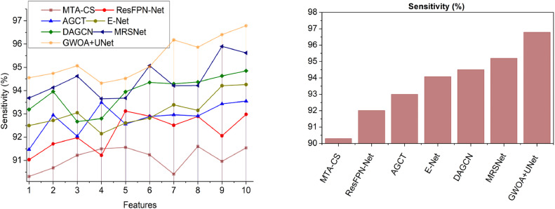
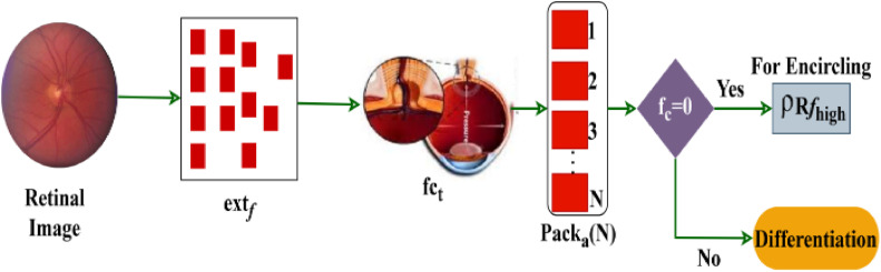
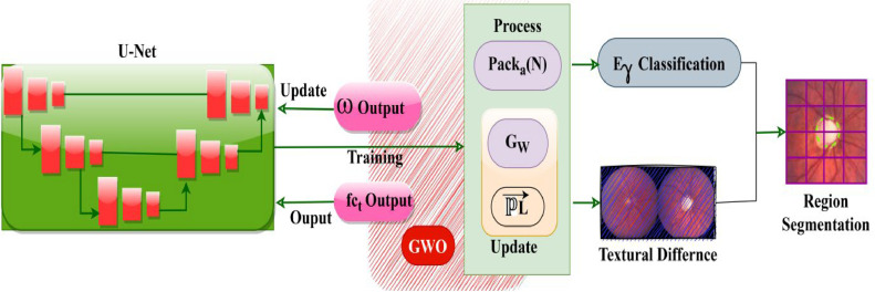
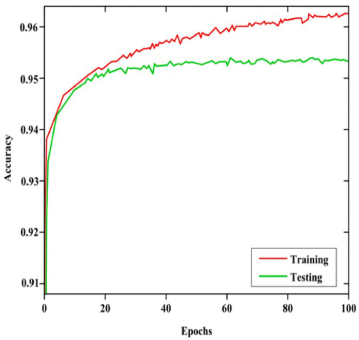
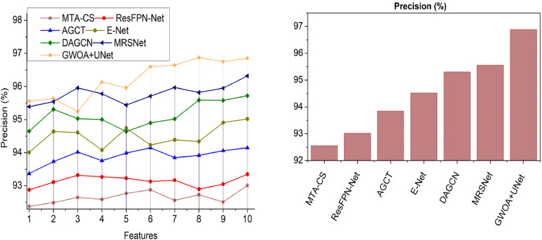
The worldwide prevalence of glaucoma makes it a major reason for blindness thus proper early diagnosis remains essential for preventing major vision deterioration. Current glaucoma screening methods that need expert handling prove to be time-intensive and complicated before yielding appropriate diagnosis and treatment. Our system addresses these difficulties through an automated glaucoma screening platform which combines advanced segmentation methods with classification approaches. A hybrid segmentation method combines Grey Wolf Optimization Algorithm with U-Shaped Networks to obtain precise extraction of the optic disc regions in retinal fundus images. Through GWOA the network achieves optimal segmentation by adopting wolf-inspired behaviors such as circular and jumping movements to identify diverse image textures. The glaucoma classification depends on CapsNet as a deep learning model that provides exceptional image detection to ensure precise diagnosis. The combination of our method delivers 96.01 % segmentation together with classification precision which outstrips traditional approaches while indicating strong capabilities for discovering glaucoma at early stages. This automated diagnosis system elevates clinical accuracy levels through an automated screening method that solves manual process limitations. The detection framework produces better accuracy to improve clinical results in a strong effort to minimize glaucoma-induced blindness worldwide and display its capabilities in real clinical environments.•Hybrid GWOA-UNet++ for precise optic disc segmentation.•CapsNet-based classification for robust glaucoma detection.•Achieved 96.01 % accuracy, surpassing existing methods.

摘要

青光眼在全球范围内的高患病率使其成为导致失明的主要原因之一,因此,早期进行正确诊断对于预防视力严重恶化至关重要。目前的青光眼筛查方法需要专家操作,在做出正确诊断和治疗之前,这些方法既耗时又复杂。我们的系统通过一个自动化青光眼筛查平台解决了这些难题,该平台将先进的分割方法与分类方法相结合。一种混合分割方法将灰狼优化算法与U型网络相结合,以精确提取眼底图像中的视盘区域。通过灰狼优化算法,该网络采用如圆周运动和跳跃运动等受狼启发的行为来识别各种图像纹理,从而实现最佳分割。青光眼分类依赖于胶囊网络(CapsNet)这一深度学习模型,该模型能提供出色的图像检测,以确保准确诊断。我们的方法相结合,实现了96.01%的分割精度和分类精度,超过了传统方法,同时显示出在早期发现青光眼的强大能力。这种自动诊断系统通过一种解决手动流程局限性的自动筛查方法,提高了临床诊断的准确性。该检测框架具有更高的准确性,以改善临床结果,努力在全球范围内将青光眼导致的失明降至最低,并在实际临床环境中展示其能力。

• 用于精确视盘分割的混合灰狼优化算法-U型网络++。

• 基于胶囊网络的分类用于可靠的青光眼检测。

• 准确率达到96.01%,超过现有方法。

https://cdn.ncbi.nlm.nih.gov/pmc/blobs/20c6/11999292/0ed90c80c15d/gr18.jpg
https://cdn.ncbi.nlm.nih.gov/pmc/blobs/20c6/11999292/75a844744c8d/ga1.jpg
https://cdn.ncbi.nlm.nih.gov/pmc/blobs/20c6/11999292/ab4c40fc1744/gr1.jpg
https://cdn.ncbi.nlm.nih.gov/pmc/blobs/20c6/11999292/6b471bc82a4b/gr2.jpg
https://cdn.ncbi.nlm.nih.gov/pmc/blobs/20c6/11999292/2f447523af5c/gr3.jpg
https://cdn.ncbi.nlm.nih.gov/pmc/blobs/20c6/11999292/bb5da8b76e5d/gr4.jpg
https://cdn.ncbi.nlm.nih.gov/pmc/blobs/20c6/11999292/d45d82127627/gr5.jpg
https://cdn.ncbi.nlm.nih.gov/pmc/blobs/20c6/11999292/660b2a1e3ae0/gr6.jpg
https://cdn.ncbi.nlm.nih.gov/pmc/blobs/20c6/11999292/e33dd5ab66f5/gr7.jpg
https://cdn.ncbi.nlm.nih.gov/pmc/blobs/20c6/11999292/4a192e15a050/gr8.jpg
https://cdn.ncbi.nlm.nih.gov/pmc/blobs/20c6/11999292/6ea3c981f740/gr9.jpg
https://cdn.ncbi.nlm.nih.gov/pmc/blobs/20c6/11999292/832d71aa5bab/gr10.jpg
https://cdn.ncbi.nlm.nih.gov/pmc/blobs/20c6/11999292/4cc5a1d25ffc/gr11.jpg
https://cdn.ncbi.nlm.nih.gov/pmc/blobs/20c6/11999292/044f767c299c/gr12.jpg
https://cdn.ncbi.nlm.nih.gov/pmc/blobs/20c6/11999292/3384387b801c/gr13.jpg
https://cdn.ncbi.nlm.nih.gov/pmc/blobs/20c6/11999292/b6652c31a660/gr14.jpg
https://cdn.ncbi.nlm.nih.gov/pmc/blobs/20c6/11999292/923e5dfad5ab/gr15.jpg
https://cdn.ncbi.nlm.nih.gov/pmc/blobs/20c6/11999292/ead86947ec3e/gr16.jpg
https://cdn.ncbi.nlm.nih.gov/pmc/blobs/20c6/11999292/480719a0b979/gr17.jpg
https://cdn.ncbi.nlm.nih.gov/pmc/blobs/20c6/11999292/0ed90c80c15d/gr18.jpg

相似文献

1
Grey wolf optimization technique with U-shaped and capsule networks-A novel framework for glaucoma diagnosis.结合U型网络和胶囊网络的灰狼优化技术——一种用于青光眼诊断的新型框架
MethodsX. 2025 Mar 31;14:103285. doi: 10.1016/j.mex.2025.103285. eCollection 2025 Jun.
2
Enhanced diabetic retinopathy detection using U-shaped network and capsule network-driven deep learning.使用U型网络和胶囊网络驱动的深度学习增强糖尿病视网膜病变检测
MethodsX. 2024 Nov 14;14:103052. doi: 10.1016/j.mex.2024.103052. eCollection 2025 Jun.
3
Enhanced Disc Herniation Classification Using Grey Wolf Optimization Based on Hybrid Feature Extraction and Deep Learning Methods.基于混合特征提取和深度学习方法的灰狼优化增强椎间盘突出分类
Tomography. 2024 Dec 26;11(1):1. doi: 10.3390/tomography11010001.
4
Capsule network-based deep learning for early and accurate diabetic retinopathy detection.基于胶囊网络的深度学习用于早期准确检测糖尿病视网膜病变
Int Ophthalmol. 2025 Feb 18;45(1):78. doi: 10.1007/s10792-024-03391-4.
5
Fully automated method for glaucoma screening using robust optic nerve head detection and unsupervised segmentation based cup-to-disc ratio computation in retinal fundus images.基于视网膜眼底图像中稳健的视神经头检测和无监督视杯/视盘比计算的青光眼筛查全自动方法。
Comput Med Imaging Graph. 2019 Oct;77:101643. doi: 10.1016/j.compmedimag.2019.101643. Epub 2019 Aug 14.
6
Rim learning framework based on TS-GAN: A new paradigm of automated glaucoma screening from fundus images.基于TS-GAN的边缘学习框架:一种从眼底图像自动进行青光眼筛查的新范式。
Comput Biol Med. 2025 Mar;187:109752. doi: 10.1016/j.compbiomed.2025.109752. Epub 2025 Feb 3.
7
ECSD-Net: A joint optic disc and cup segmentation and glaucoma classification network based on unsupervised domain adaptation.ECSD-Net:一种基于无监督域自适应的联合视盘和杯分割及青光眼分类网络。
Comput Methods Programs Biomed. 2022 Jan;213:106530. doi: 10.1016/j.cmpb.2021.106530. Epub 2021 Nov 14.
8
An optimized two stage U-Net approach for segmentation of pancreas and pancreatic tumor.一种用于胰腺和胰腺肿瘤分割的优化两阶段U-Net方法。
MethodsX. 2024 Oct 4;13:102995. doi: 10.1016/j.mex.2024.102995. eCollection 2024 Dec.
9
Automated glaucoma diagnosis: Optimized hybrid classification model with improved U-net segmentation.自动化青光眼诊断:具有改进型U-net分割的优化混合分类模型。
Network. 2025 Mar 27:1-55. doi: 10.1080/0954898X.2025.2481958.
10
Optimized chest X-ray image semantic segmentation networks for COVID-19 early detection.用于 COVID-19 早期检测的优化胸部 X 射线图像语义分割网络。
J Xray Sci Technol. 2022;30(3):491-512. doi: 10.3233/XST-211113.

本文引用的文献

1
Enhanced leukemia prediction using hybrid ant colony and ant lion optimization for gene selection and classification.使用混合蚁群和蚁狮优化进行基因选择与分类以增强白血病预测
MethodsX. 2025 Feb 20;14:103239. doi: 10.1016/j.mex.2025.103239. eCollection 2025 Jun.
2
Capsule network-based deep learning for early and accurate diabetic retinopathy detection.基于胶囊网络的深度学习用于早期准确检测糖尿病视网膜病变
Int Ophthalmol. 2025 Feb 18;45(1):78. doi: 10.1007/s10792-024-03391-4.
3
Enhancing glaucoma diagnosis: Generative adversarial networks in synthesized imagery and classification with pretrained MobileNetV2.
增强青光眼诊断:合成图像中的生成对抗网络以及使用预训练的MobileNetV2进行分类
MethodsX. 2024 Dec 18;14:103116. doi: 10.1016/j.mex.2024.103116. eCollection 2025 Jun.
4
Enhanced diabetic retinopathy detection using U-shaped network and capsule network-driven deep learning.使用U型网络和胶囊网络驱动的深度学习增强糖尿病视网膜病变检测
MethodsX. 2024 Nov 14;14:103052. doi: 10.1016/j.mex.2024.103052. eCollection 2025 Jun.
5
The Application of Deep Learning for the Segmentation and Classification of Coronary Arteries.深度学习在冠状动脉分割与分类中的应用
Diagnostics (Basel). 2023 Jul 5;13(13):2274. doi: 10.3390/diagnostics13132274.
6
Automatic Diagnosis of Glaucoma from Retinal Images Using Deep Learning Approach.使用深度学习方法从视网膜图像中自动诊断青光眼
Diagnostics (Basel). 2023 May 14;13(10):1738. doi: 10.3390/diagnostics13101738.
7
Automatic Detection of Microaneurysms in Fundus Images Using an Ensemble-Based Segmentation Method.基于集成的眼底图像微动脉瘤自动检测分割方法。
Sensors (Basel). 2023 Mar 24;23(7):3431. doi: 10.3390/s23073431.
8
Dual Consistency Enabled Weakly and Semi-Supervised Optic Disc and Cup Segmentation With Dual Adaptive Graph Convolutional Networks.基于双自适应图卷积网络的双一致性弱监督和半监督视盘与视杯分割
IEEE Trans Med Imaging. 2023 Feb;42(2):416-429. doi: 10.1109/TMI.2022.3203318. Epub 2023 Feb 2.
9
Deep learning-based medical image segmentation of the aorta using XR-MSF-U-Net.基于深度学习的 XR-MSF-U-Net 主动脉医学图像分割。
Comput Methods Programs Biomed. 2022 Oct;225:107073. doi: 10.1016/j.cmpb.2022.107073. Epub 2022 Aug 11.
10
Classification of retinal images based on convolutional neural network.基于卷积神经网络的视网膜图像分类
Microsc Res Tech. 2021 Mar;84(3):394-414. doi: 10.1002/jemt.23596. Epub 2020 Dec 22.