Tang Jack C, Pan Dorothy W, Oghalai John S, Applegate Brian E
University of Southern California, Caruso Department of Otolaryngology-Head and Neck Surgery, Los Angeles, California, United States.
University of Southern California, Alfred Mann Department of Biomedical Engineering, Los Angeles, California, United States.
J Biomed Opt. 2025 Apr;30(4):046007. doi: 10.1117/1.JBO.30.4.046007. Epub 2025 Apr 17.
There is no clinical imaging method to visualize the soft tissues of the human cochlea, which are crucial for sound transduction and are damaged in sensorineural hearing loss. Although optical coherence tomography (OCT) has been effective in small animal models, we show for the first time that it can image through the full thickness of the human otic capsule and resolve cochlear microstructures despite increased scattering.
We aim to investigate whether OCT could image the cochlea through the otic capsule. We compared 1.7 and OCT to test if the reduced scattering at provided any appreciable advantage for imaging the cochleae.
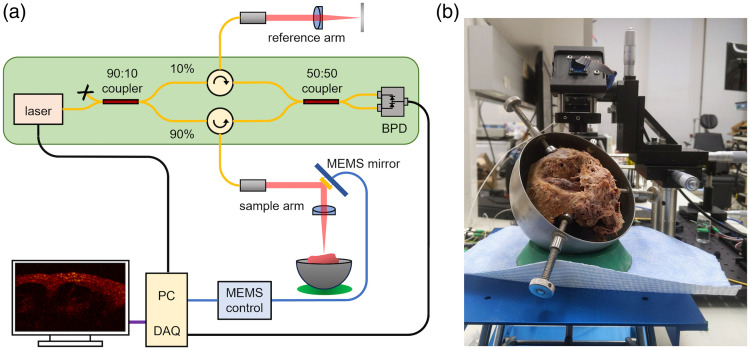
OCT interferometers were built for both 1.3 and wavelengths, using identical sample and reference arm optics in both systems. Imaging was performed on two fixed human temporal bones with intact cochleae. The interferometers were designed to allow seamless switching between 1.3 and OCT without disrupting the temporal bone during imaging.
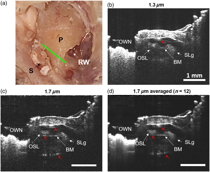
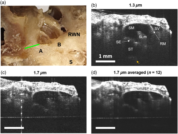
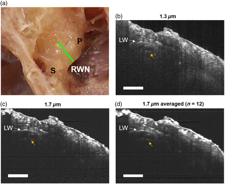
We took volumetric OCT images at the base, apex, and hook regions of fixed human cochleae and compared the images taken at with those taken at . At both wavelengths, we could see through the otic capsule and identify cochlear structures. In some cases, OCT resulted in clearer images of the lateral wall, interior scala, and fine cochlear structures due to reduced multiple scattering at depth compared with .
We conclude that both and OCT can image through the human otic capsule, offering the potential for direct measurement of cochlear vibrometry or blood flow in living humans. Using light, we observed reduced multiple scattering in the otic capsule, leading to enhanced contrast of cochlear structures compared with . However, these improvements were marginal and came with trade-offs.
目前尚无临床成像方法能够可视化人类耳蜗的软组织,而这些软组织对于声音传导至关重要,且在感音神经性听力损失中会受到损害。尽管光学相干断层扫描(OCT)在小动物模型中已取得成效,但我们首次证明,尽管散射增加,它仍能穿透人类听骨囊的全层进行成像,并分辨出耳蜗的微观结构。
我们旨在研究OCT是否能够透过听骨囊对耳蜗进行成像。我们比较了1.7微米和 微米的OCT,以测试在 微米波长下散射减少是否为耳蜗成像带来显著优势。
针对1.3微米和 微米波长构建了OCT干涉仪,两个系统均使用相同的样品臂和参考臂光学元件。对两只耳蜗完整的固定人类颞骨进行成像。干涉仪设计为能够在1.3微米和 微米的OCT之间无缝切换,且在成像过程中不会干扰颞骨。
我们获取了固定人类耳蜗基部、顶部和钩部区域的OCT体积图像,并将在 微米波长下拍摄的图像与在 微米波长下拍摄的图像进行比较。在两个波长下,我们都能够透过听骨囊并识别耳蜗结构。在某些情况下,与 微米波长相比, 微米波长的OCT由于深度处多次散射减少,使得侧壁、内耳道和精细耳蜗结构的图像更清晰。
我们得出结论,1.3微米和 微米的OCT均能透过人类听骨囊进行成像,为在活体人类中直接测量耳蜗振动或血流提供了可能性。使用 微米波长的光,我们观察到听骨囊中多次散射减少,与 微米波长相比,耳蜗结构的对比度增强。然而,这些改进很有限,且存在权衡。