Garavelli Chiara, Aldieri Alessandra, Palanca Marco, Dall'Ara Enrico, Viceconti Marco
Department of Industrial Engineering, Alma Mater Studiorum - University of Bologna, Bologna, Italy.
Medical Technology Lab, IRCCS Istituto Ortopedico Rizzoli, Bologna, Italy.
Biomech Model Mechanobiol. 2025 Jun;24(3):1017-1030. doi: 10.1007/s10237-025-01950-x. Epub 2025 Apr 19.
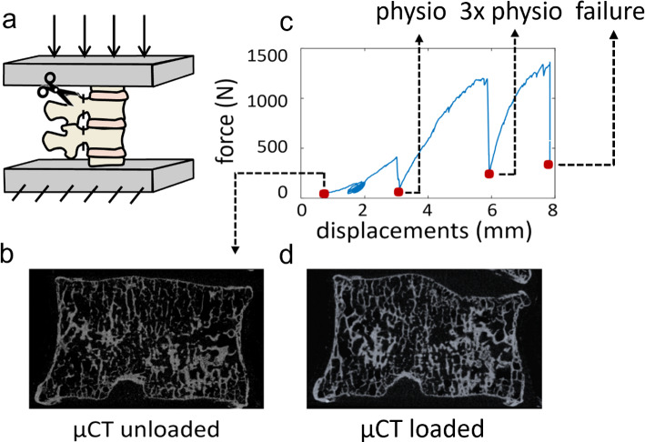
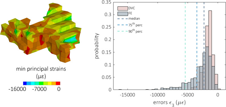
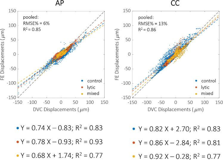
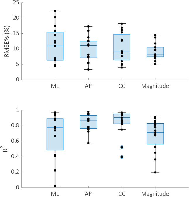
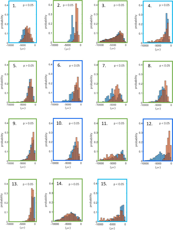
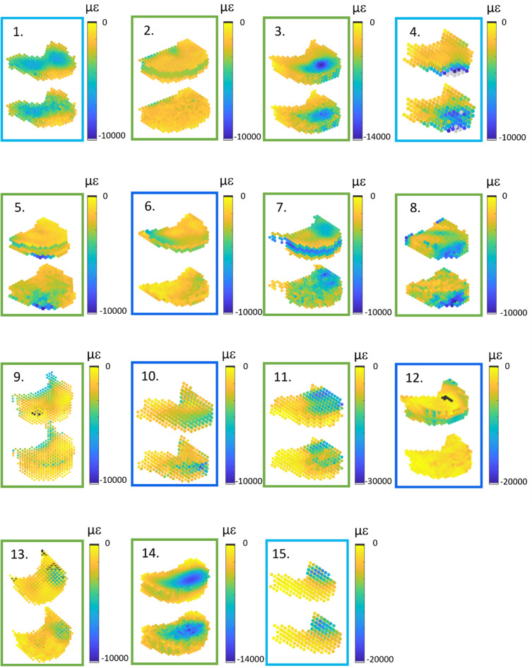
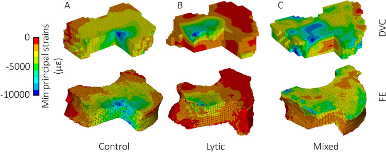
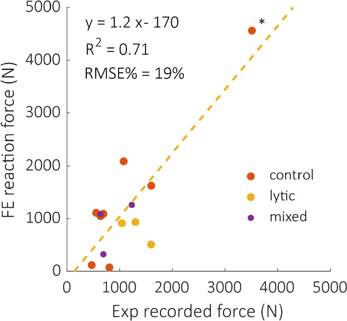
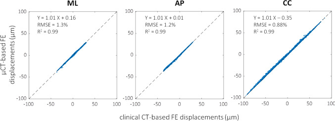
Several conditions can increase the incidence of vertebral fragility fractures, including metastatic bone disease. Computational tools could help clinicians estimate the risk of vertebral fracture in these patients; however, comparison with in vitro data is mandatory before using them in clinical practice. Nine spine segments were tested under compression and imaged with micro-computed tomography (µCT). The displacement field was calculated for each vertebra using a global digital volume correlation (DVC) approach. Subject-specific homogenised finite element models of each vertebra were built from µCT images, applying experimentally matched boundary conditions at the endplates. Numerical and experimental displacements, reaction forces, and locations showing higher strain concentrations were eventually compared. Additionally, given that µCT cannot be performed in clinical settings, the outcomes of a µCT-based model were also compared to those of a model built from clinical CT scans of the same specimen. Good agreement between DVC and µCT-based FE displacements was found, both for healthy (R = 0.69 ÷ 0.83, RMSE = 3 ÷ 22%, max error < 45 μm) and metastatic (R = 0.64 ÷ 0.93, RMSE = 5 ÷ 18%, max error < 54 μm) vertebrae. Strong correlations were found between µCT-based and clinical CT-based FE model outcomes (R = 0.99, RMSE < 1.3%, max difference = 6 μm). Furthermore, the models qualitatively identified the most deformed regions identified with the experiments. In conclusion, the combination of experimental full-field technique and in-silico modelling enabled the development of a promising pipeline to validate bone strength predictors in the elastic range. Further improvements are needed to analyse vertebral post-yield behaviour better.
几种情况会增加椎体脆性骨折的发生率,包括转移性骨病。计算工具可帮助临床医生评估这些患者发生椎体骨折的风险;然而,在临床实践中使用这些工具之前,必须与体外数据进行比较。对九个脊柱节段进行了压缩测试,并用微型计算机断层扫描(µCT)成像。使用全局数字体积相关(DVC)方法计算每个椎体的位移场。根据µCT图像建立每个椎体的特定受试者均质有限元模型,并在终板处应用实验匹配的边界条件。最终比较了数值和实验位移、反作用力以及显示较高应变集中的位置。此外,鉴于在临床环境中无法进行µCT检查,还将基于µCT的模型结果与从同一标本的临床CT扫描建立的模型结果进行了比较。在健康椎体(R = 0.69÷0.83,RMSE = 3÷22%,最大误差<45μm)和转移性椎体(R = 0.64÷0.93,RMSE = 5÷18%,最大误差<54μm)中,DVC与基于µCT的有限元位移之间均发现了良好的一致性。基于µCT的有限元模型结果与基于临床CT的有限元模型结果之间发现了强相关性(R = 0.99,RMSE<1.3%,最大差异 = 6μm)。此外,这些模型定性地识别出了实验中确定的变形最大的区域。总之,实验全场技术和计算机模拟建模的结合使得开发出一种有前景的流程成为可能,以验证弹性范围内的骨强度预测指标。需要进一步改进以更好地分析椎体屈服后行为。