Cheheltani Sepideh, Islam Sadia T, Malino Heather, Abera Kalekidan, Aryal Sandeep, Forbes Karen, Parreno Justin, Fowler Velia M
Department of Biological Sciences, University of Delaware, Newark, DE, United States.
Department of Biomedical Engineering, University of Delaware, Newark, DE, United States.
Front Ophthalmol (Lausanne). 2025 Apr 4;5:1562583. doi: 10.3389/fopht.2025.1562583. eCollection 2025.
Proper ocular lens function requires biomechanical flexibility, which is reduced during aging. As increasing lens size has been shown to correlate with lens biomechanical stiffness in aging, we tested the hypothesis that whole lens size determines gross biomechanical stiffness by comparing lenses of varying sizes from three rodent species (mice, rats, and guinea pigs).
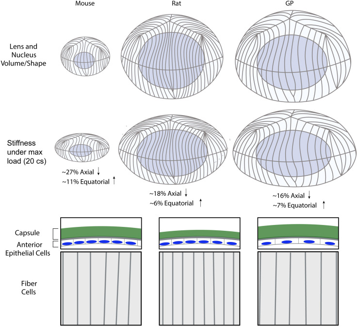
Coverslip compression assay was performed to measure whole lens biomechanics. Whole mount staining on fixed lenses, followed by confocal microscopy, was conducted to measure lens microstructures.
Among the three species, guinea pig lenses are the largest, rat lenses are smaller than guinea pig lenses, and mouse lenses are the smallest of the three. We found that rat and guinea pig lenses are stiffer than the much smaller mouse lenses. However, despite guinea pig lenses being larger than rat lenses, whole lens stiffness between guinea pigs and rats is not different. This refutes our hypothesis and indicates that lens size does not solely determine lens stiffness. We next compared lens microstructures, including nuclear size, capsule thickness, epithelial cell area, fiber cell widths, and suture organization between mice, rats, and guinea pigs. The lens nucleus is the largest in guinea pigs, followed by rats, and mice. However, the rat nucleus occupies a larger fraction of the lens. Both lens capsule thickness and fiber cell widths are the largest in guinea pigs, followed by mice and then rats. Epithelial cells are the largest in guinea pigs, and there are no differences between mice and rats. In addition, the lens suture shape appears similar across all three species.
Overall, our data indicates that whole lens size and microstructure morphometrics do not correlate with lens stiffness, indicating that factors contributing to lens biomechanics are complex and likely multifactorial.
晶状体正常功能需要生物力学灵活性,而这种灵活性在衰老过程中会降低。由于研究表明晶状体尺寸增加与衰老过程中晶状体生物力学硬度相关,我们通过比较三种啮齿动物(小鼠、大鼠和豚鼠)不同大小的晶状体,来检验整个晶状体大小决定总体生物力学硬度这一假设。
进行盖玻片压缩试验以测量整个晶状体的生物力学。对固定的晶状体进行整装染色,然后用共聚焦显微镜测量晶状体微观结构。
在这三个物种中,豚鼠晶状体最大,大鼠晶状体比豚鼠晶状体小,小鼠晶状体是三者中最小的。我们发现大鼠和豚鼠的晶状体比小得多的小鼠晶状体更硬。然而,尽管豚鼠晶状体比大鼠晶状体大,但豚鼠和大鼠之间整个晶状体的硬度并无差异。这反驳了我们的假设,表明晶状体大小并非唯一决定晶状体硬度的因素。接下来,我们比较了小鼠、大鼠和豚鼠之间晶状体的微观结构,包括核大小、囊膜厚度、上皮细胞面积、纤维细胞宽度和缝线结构。晶状体核在豚鼠中最大,其次是大鼠,小鼠最小。然而,大鼠的晶状体核在晶状体中所占比例更大。晶状体囊膜厚度和纤维细胞宽度在豚鼠中都是最大的,其次是小鼠,然后是大鼠。上皮细胞在豚鼠中最大,小鼠和大鼠之间没有差异。此外,所有三个物种的晶状体缝线形状看起来相似。
总体而言,我们的数据表明整个晶状体大小和微观结构形态测量与晶状体硬度不相关,这表明影响晶状体生物力学的因素很复杂,可能是多因素的。