• 文献检索
  • 文档翻译
  • 深度研究
  • 学术资讯
  • Suppr Zotero 插件Zotero 插件
  • 邀请有礼
  • 套餐&价格
  • 历史记录
应用&插件
Suppr Zotero 插件Zotero 插件浏览器插件Mac 客户端Windows 客户端微信小程序
定价
高级版会员购买积分包购买API积分包
服务
文献检索文档翻译深度研究API 文档MCP 服务
关于我们
关于 Suppr公司介绍联系我们用户协议隐私条款
关注我们

Suppr 超能文献

核心技术专利:CN118964589B侵权必究
粤ICP备2023148730 号-1Suppr @ 2026

文献检索

告别复杂PubMed语法,用中文像聊天一样搜索,搜遍4000万医学文献。AI智能推荐,让科研检索更轻松。

立即免费搜索

文件翻译

保留排版,准确专业,支持PDF/Word/PPT等文件格式,支持 12+语言互译。

免费翻译文档

深度研究

AI帮你快速写综述,25分钟生成高质量综述,智能提取关键信息,辅助科研写作。

立即免费体验

EphA2 和 Ephrin-A5 指导眼睛晶状体缝线对准并影响整个晶状体的弹性。

EphA2 and Ephrin-A5 Guide Eye Lens Suture Alignment and Influence Whole Lens Resilience.

机构信息

School of Optometry and Vision Science Program, Indiana University, Bloomington, Indiana, United States.

出版信息

Invest Ophthalmol Vis Sci. 2021 Dec 1;62(15):3. doi: 10.1167/iovs.62.15.3.

DOI:10.1167/iovs.62.15.3
PMID:34854885
原文链接:https://pmc.ncbi.nlm.nih.gov/articles/PMC8648058/
Abstract

PURPOSE

Fine focusing of light by the eye lens onto the retina relies on the ability of the lens to change shape during the process of accommodation. Little is known about the cellular structures that regulate elasticity and resilience. We tested whether Eph-ephrin signaling is involved in lens biomechanical properties.

METHODS

We used confocal microscopy and tissue mechanical testing to examine mouse lenses with genetic disruption of EphA2 or ephrin-A5.

RESULTS

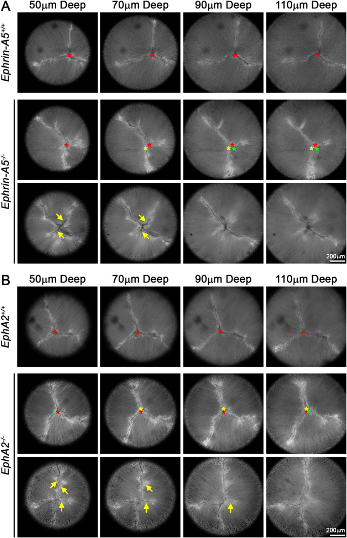
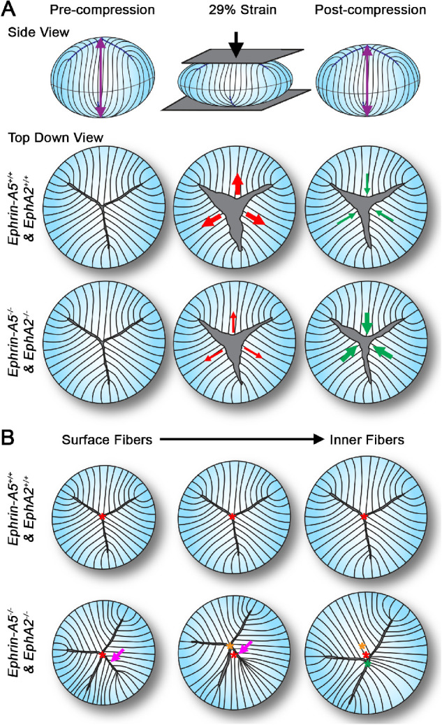
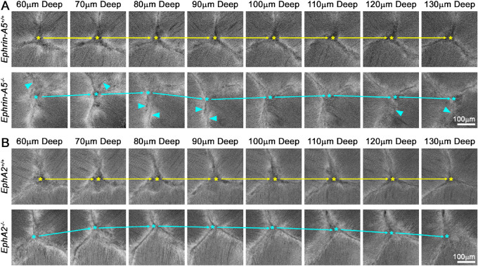
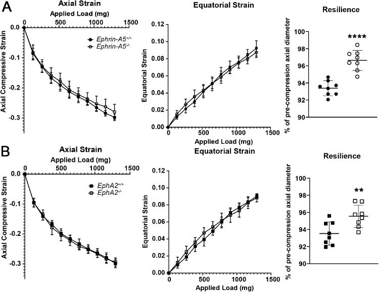
Confocal imaging revealed misalignment of the suture between each shell of newly added fiber cells in knockout lenses. Despite having disordered sutures, loss of EphA2 or ephrin-A5 did not affect lens stiffness. Surprisingly, knockout lenses were more resilient and recovered almost completely after load removal. Confocal microscopy and quantitative image analysis from live lenses before, during, and after compression revealed that knockout lenses had misaligned Y-sutures, leading to a change in force distribution during compression. Knockout lenses displayed decreased separation of fiber cell tips at the anterior suture at high loads and had more complete recovery after load removal, which leads to improved whole-lens resiliency.

CONCLUSIONS

EphA2 and ephrin-A5 are needed for normal patterning of fiber cell tips and the formation of a well-aligned Y-suture with fiber tips stacked on top of previous generations of fiber cells. The misalignment of lens sutures leads to increased resilience after compression. The data suggest that alignment of the Y-suture may constrain the overall elasticity and resilience of the lens.

摘要

目的

眼睛晶状体将光线精确定焦在视网膜上依赖于晶状体在调节过程中改变形状的能力。对于调节弹性和弹性恢复的细胞结构知之甚少。我们测试了 Eph-ephrin 信号是否参与晶状体生物力学特性。

方法

我们使用共聚焦显微镜和组织力学测试来检查 EphA2 或 ephrin-A5 基因敲除的小鼠晶状体。

结果

共聚焦成像显示,在敲除的晶状体中,每个新添加的纤维细胞壳之间的缝线不对齐。尽管缝线排列紊乱,但 EphA2 或 ephrin-A5 的缺失并不影响晶状体的硬度。令人惊讶的是,敲除的晶状体更有弹性,在负载移除后几乎完全恢复。在压缩前、压缩中和压缩后对活体晶状体进行共聚焦显微镜和定量图像分析显示,敲除的晶状体的 Y 缝线不对齐,导致在压缩过程中力分布发生变化。在高负荷下,敲除的晶状体的前部缝线处纤维细胞尖端分离减少,在负载移除后恢复更完全,从而提高了整个晶状体的弹性。

结论

EphA2 和 ephrin-A5 对于纤维细胞尖端的正常排列以及形成与纤维尖端堆叠在上一代纤维细胞之上的对齐良好的 Y 缝线是必需的。晶状体缝线的不对齐导致压缩后弹性增加。数据表明,Y 缝线的对齐可能会限制晶状体的整体弹性和弹性恢复。

https://cdn.ncbi.nlm.nih.gov/pmc/blobs/dc13/8648058/27175031f838/iovs-62-15-3-f005.jpg
https://cdn.ncbi.nlm.nih.gov/pmc/blobs/dc13/8648058/adaabc39c105/iovs-62-15-3-f001.jpg
https://cdn.ncbi.nlm.nih.gov/pmc/blobs/dc13/8648058/7d7478cdac79/iovs-62-15-3-f002.jpg
https://cdn.ncbi.nlm.nih.gov/pmc/blobs/dc13/8648058/a98b6a6b4150/iovs-62-15-3-f003.jpg
https://cdn.ncbi.nlm.nih.gov/pmc/blobs/dc13/8648058/cd66f5c22313/iovs-62-15-3-f004.jpg
https://cdn.ncbi.nlm.nih.gov/pmc/blobs/dc13/8648058/27175031f838/iovs-62-15-3-f005.jpg

相似文献

1
EphA2 and Ephrin-A5 Guide Eye Lens Suture Alignment and Influence Whole Lens Resilience.EphA2 和 Ephrin-A5 指导眼睛晶状体缝线对准并影响整个晶状体的弹性。
Invest Ophthalmol Vis Sci. 2021 Dec 1;62(15):3. doi: 10.1167/iovs.62.15.3.
2
EphA2 and ephrin-A5 are not a receptor-ligand pair in the ocular lens.EphA2和ephrin-A5在晶状体中并非受体-配体对。
Exp Eye Res. 2017 Sep;162:9-17. doi: 10.1016/j.exer.2017.06.016. Epub 2017 Jun 23.
3
EphA2 Affects Development of the Eye Lens Nucleus and the Gradient of Refractive Index.EphA2 影响眼睛晶状体核的发育和折射率梯度。
Invest Ophthalmol Vis Sci. 2022 Jan 3;63(1):2. doi: 10.1167/iovs.63.1.2.
4
Diverse roles of Eph/ephrin signaling in the mouse lens.Eph/ephrin 信号在小鼠晶状体中的多种作用。
PLoS One. 2011;6(11):e28147. doi: 10.1371/journal.pone.0028147. Epub 2011 Nov 29.
5
Loss of ephrin-A5 function disrupts lens fiber cell packing and leads to cataract.Ephrin-A5功能丧失会破坏晶状体纤维细胞的排列并导致白内障。
Proc Natl Acad Sci U S A. 2008 Oct 28;105(43):16620-5. doi: 10.1073/pnas.0808987105. Epub 2008 Oct 23.
6
Mapping the Universe of Eph Receptor and Ephrin Ligand Transcripts in Epithelial and Fiber Cells of the Eye Lens.绘制眼部晶状体上皮细胞和纤维细胞中 Eph 受体和 Ephrin 配体转录本的图谱。
Cells. 2022 Oct 19;11(20):3291. doi: 10.3390/cells11203291.
7
Epha2 and Efna5 participate in lens cell pattern-formation.Epha2 和 Efna5 参与晶状体细胞的形态发生。
Differentiation. 2018 Jul-Aug;102:1-9. doi: 10.1016/j.diff.2018.05.002. Epub 2018 May 17.
8
Roles of Eph-Ephrin Signaling in the Eye Lens Cataractogenesis, Biomechanics, and Homeostasis.Eph-Ephrin信号通路在晶状体白内障形成、生物力学及内环境稳定中的作用
Front Cell Dev Biol. 2022 Feb 28;10:852236. doi: 10.3389/fcell.2022.852236. eCollection 2022.
9
Eph-ephrin Signaling Affects Eye Lens Fiber Cell Intracellular Voltage and Membrane Conductance.Eph-ephrin信号传导影响眼晶状体纤维细胞的细胞内电压和膜电导。
Front Physiol. 2021 Nov 25;12:772276. doi: 10.3389/fphys.2021.772276. eCollection 2021.
10
Further analysis of the lens of ephrin-A5-/- mice: development of postnatal defects.对ephrin-A5基因敲除小鼠晶状体的进一步分析:产后缺陷的发展。
Mol Vis. 2013;19:254-66. Epub 2013 Feb 3.

引用本文的文献

1
Comparative analysis of rodent lens morphometrics and biomechanical properties.啮齿动物晶状体形态测量与生物力学特性的比较分析。
Front Ophthalmol (Lausanne). 2025 Apr 4;5:1562583. doi: 10.3389/fopht.2025.1562583. eCollection 2025.
2
Canonical ligand-dependent and non-canonical ligand-independent EphA2 signaling in the eye lens of wild-type, knockout, and aging mice.野生型、敲除型和衰老型小鼠眼睛晶状体中的经典配体依赖性和非经典配体非依赖性 EphA2 信号传导。
Aging (Albany NY). 2024 Oct 25;16(20):13039-13075. doi: 10.18632/aging.206144.
3
Tissue, cellular, and molecular level determinants for eye lens stiffness and elasticity.

本文引用的文献

1
Age-related changes in eye lens biomechanics, morphology, refractive index and transparency.晶状体生物力学、形态、折射率和透明度的年龄相关变化。
Aging (Albany NY). 2019 Dec 16;11(24):12497-12531. doi: 10.18632/aging.102584.
2
Tropomyosin 3.5 protects the F-actin networks required for tissue biomechanical properties.原肌球蛋白 3.5 保护组织生物力学特性所需的 F-肌动蛋白网络。
J Cell Sci. 2018 Nov 29;131(23):jcs222042. doi: 10.1242/jcs.222042.
3
The effects of mechanical strain on mouse eye lens capsule and cellular microstructure.
晶状体硬度和弹性的组织、细胞及分子水平决定因素。
Front Ophthalmol (Lausanne). 2024 Aug 8;4:1456474. doi: 10.3389/fopht.2024.1456474. eCollection 2024.
4
The significance of growth shells in development of symmetry, transparency, and refraction of the human lens.生长线在人晶状体对称性、透明度和屈光发育中的意义。
Front Ophthalmol (Lausanne). 2024 Jul 19;4:1434327. doi: 10.3389/fopht.2024.1434327. eCollection 2024.
5
Spatial-temporal comparison of Eph/Ephrin gene expression in ocular lenses from aging and knockout mice.衰老和基因敲除小鼠晶状体中Eph/Ephrin基因表达的时空比较。
Front Ophthalmol (Lausanne). 2024 Jun 4;4:1410860. doi: 10.3389/fopht.2024.1410860. eCollection 2024.
6
Disease-related non-muscle myosin IIA D1424N rod domain mutation, but not R702C motor domain mutation, disrupts mouse ocular lens fiber cell alignment and hexagonal packing.疾病相关的非肌肉肌球蛋白IIA D1424N杆状结构域突变,而非R702C运动结构域突变,破坏了小鼠眼晶状体纤维细胞的排列和六边形堆积。
Cytoskeleton (Hoboken). 2024 Dec;81(12):789-805. doi: 10.1002/cm.21853. Epub 2024 Mar 22.
7
Spatially Resolved Proteomics Reveals Lens Suture-Related Cell-Cell Junctional Protein Distributions.空间分辨蛋白质组学揭示晶状体缝合线相关细胞-细胞连接蛋白的分布。
Invest Ophthalmol Vis Sci. 2023 Aug 1;64(11):28. doi: 10.1167/iovs.64.11.28.
8
Adaptive Optical Two-Photon Fluorescence Microscopy Probes Cellular Organization of Ocular Lenses In Vivo.自适应光学双光子荧光显微镜探测活体眼晶状体的细胞结构。
Invest Ophthalmol Vis Sci. 2023 Jun 1;64(7):20. doi: 10.1167/iovs.64.7.20.
9
Nonmuscle Myosin IIA Regulates the Precise Alignment of Hexagonal Eye Lens Epithelial Cells During Fiber Cell Formation and Differentiation.非肌肉肌球蛋白 IIA 调控六边形眼晶状体上皮细胞在纤维细胞形成和分化过程中的精确排列。
Invest Ophthalmol Vis Sci. 2023 Apr 3;64(4):20. doi: 10.1167/iovs.64.4.20.
10
Adaptive optical two-photon fluorescence microscopy probes cellular organization of ocular lenses in vivo.自适应光学双光子荧光显微镜可在体内探测晶状体的细胞组织结构。
bioRxiv. 2023 Jan 19:2023.01.17.524320. doi: 10.1101/2023.01.17.524320.
机械应变对小鼠眼晶状体囊和细胞微观结构的影响。
Mol Biol Cell. 2018 Aug 8;29(16):1963-1974. doi: 10.1091/mbc.E18-01-0035. Epub 2018 Apr 10.
4
Epha2 and Efna5 participate in lens cell pattern-formation.Epha2 和 Efna5 参与晶状体细胞的形态发生。
Differentiation. 2018 Jul-Aug;102:1-9. doi: 10.1016/j.diff.2018.05.002. Epub 2018 May 17.
5
EphA2 and ephrin-A5 are not a receptor-ligand pair in the ocular lens.EphA2和ephrin-A5在晶状体中并非受体-配体对。
Exp Eye Res. 2017 Sep;162:9-17. doi: 10.1016/j.exer.2017.06.016. Epub 2017 Jun 23.
6
Tropomodulin 1 Regulation of Actin Is Required for the Formation of Large Paddle Protrusions Between Mature Lens Fiber Cells.原肌球蛋白1对肌动蛋白的调节是成熟晶状体纤维细胞之间形成大型桨状突起所必需的。
Invest Ophthalmol Vis Sci. 2016 Aug 1;57(10):4084-99. doi: 10.1167/iovs.16-19949.
7
Sequential Application of Glass Coverslips to Assess the Compressive Stiffness of the Mouse Lens: Strain and Morphometric Analyses.连续应用盖玻片评估小鼠晶状体的压缩刚度:应变和形态计量分析。
J Vis Exp. 2016 May 3(111):53986. doi: 10.3791/53986.
8
Ephrin-A2 and ephrin-A5 guide contralateral targeting but not topographic mapping of ventral cochlear nucleus axons.埃菲林-A2和埃菲林-A5引导对侧靶向,但不引导腹侧耳蜗核轴突的拓扑映射。
Neural Dev. 2015 Dec 15;10:27. doi: 10.1186/s13064-015-0054-6.
9
Breakdown of interlocking domains may contribute to formation of membranous globules and lens opacity in ephrin-A5(-/-) mice.在ephrin - A5基因敲除小鼠中,连锁结构域的破坏可能导致膜性小球的形成和晶状体混浊。
Exp Eye Res. 2016 Apr;145:130-139. doi: 10.1016/j.exer.2015.11.017. Epub 2015 Nov 28.
10
Ankyrin-B directs membrane tethering of periaxin and is required for maintenance of lens fiber cell hexagonal shape and mechanics.锚蛋白B指导外周蛋白的膜连接,并且是维持晶状体纤维细胞六边形形状和力学特性所必需的。
Am J Physiol Cell Physiol. 2016 Jan 15;310(2):C115-26. doi: 10.1152/ajpcell.00111.2015. Epub 2015 Nov 4.