• 文献检索
  • 文档翻译
  • 深度研究
  • 学术资讯
  • Suppr Zotero 插件Zotero 插件
  • 邀请有礼
  • 套餐&价格
  • 历史记录
应用&插件
Suppr Zotero 插件Zotero 插件浏览器插件Mac 客户端Windows 客户端微信小程序
定价
高级版会员购买积分包购买API积分包
服务
文献检索文档翻译深度研究API 文档MCP 服务
关于我们
关于 Suppr公司介绍联系我们用户协议隐私条款
关注我们

Suppr 超能文献

核心技术专利:CN118964589B侵权必究
粤ICP备2023148730 号-1Suppr @ 2026

文献检索

告别复杂PubMed语法,用中文像聊天一样搜索,搜遍4000万医学文献。AI智能推荐,让科研检索更轻松。

立即免费搜索

文件翻译

保留排版,准确专业,支持PDF/Word/PPT等文件格式,支持 12+语言互译。

免费翻译文档

深度研究

AI帮你快速写综述,25分钟生成高质量综述,智能提取关键信息,辅助科研写作。

立即免费体验

通过表面肌电图的变分模态分解识别和量化用于踝关节康复的肌肉共同收缩。

Identification and quantification of muscular cocontraction for ankle rehabilitation through variational mode decomposition in surface electromyography.

作者信息

Yasmeen Sania, Waris Asim, Amin Faisal, Iqbal Javaid, Gilani Syed Omer, Khan Muhammad Jawad, Hazzazi Fawwaz, Imran Ahmed, Shah Umer Hameed, Ijaz Muhammad Adeel

机构信息

Department of Biomedical Engineering and Sciences, School of Mechanical and Manufacturing Engineering, National University of Sciences and Technology (NUST), Islamabad, Pakistan.

Department of Electrical, Computer, and Biomedical Engineering, Abu Dhabi University, Abu Dhabi, UAE.

出版信息

Sci Rep. 2025 Apr 28;15(1):14847. doi: 10.1038/s41598-025-96334-7.

DOI:10.1038/s41598-025-96334-7
PMID:40295627
原文链接:https://pmc.ncbi.nlm.nih.gov/articles/PMC12037877/
Abstract

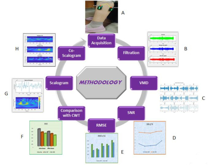
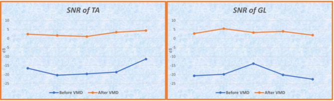
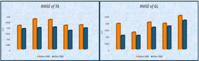
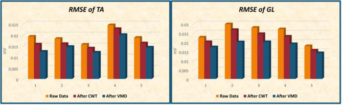
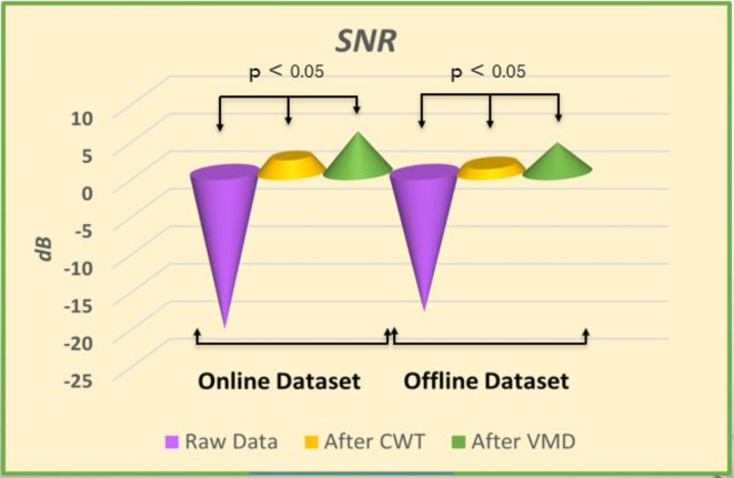
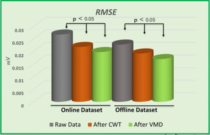
The ankle joint plays important role in performing fundamental activities such as walking and other essential daily tasks. Ankle stabilization and muscle co-contraction are crucial for rehabilitating gait abnormalities, as impaired ankle function disrupts gait, causes pain and inflammation, and hampers recovery. Accurate assessment of muscle co-contraction is crucial for developing effective non-pharmacological interventions. This paper introduces a novel approach using Variational Mode Decomposition (VMD) combined with scalogram visualization technique to analyze surface electromyographic (sEMG) signals from antagonist muscles of the lower limb and assesses muscular co-contraction using the coscalogram function. The present study compares VMD with the Continuous Wavelet Transform (CWT) approach and shows that VMD outperforms CWT in terms of both SNR and RMSE. On average, the increase in SNR in case of VMD (-17.65 ± 8.1dB to 2.98 ± 2.2dB) was greater than that of CWT (-17.65 ± 3.7dB to 1.34 ± 1.5dB). Similarly, the reduction in RMSE with VMD (0.023 ± 0.0029 to 0.017 ± 0.0015) surpassed that achieved with CWT (0.023 ± 0.0027 to 0.020 ± 0.0025). This enhanced accuracy in identifying co-contraction events has the potential to significantly improve clinical assessment and rehabilitation strategies for patients with ankle joint pathologies. To further validate VMD's effectiveness, we quantitatively assessed co-contraction events by comparing mean peak amplitudes identified using VMD and CWT. Our analysis, which revealed that VMD consistently captured stronger co-contraction events (higher mean peak amplitudes), supports VMD's superiority in accurately identifying and quantifying ankle muscle co-contraction. These results have significant implications for clinical practice, offering the potential for more precise assessments of ankle joint function and the development of more targeted and effective rehabilitation interventions.

摘要

踝关节在执行诸如行走和其他基本日常任务等基本活动中发挥着重要作用。踝关节稳定和肌肉协同收缩对于恢复步态异常至关重要,因为踝关节功能受损会扰乱步态、导致疼痛和炎症,并阻碍恢复。准确评估肌肉协同收缩对于开发有效的非药物干预措施至关重要。本文介绍了一种使用变分模态分解(VMD)结合小波尺度图可视化技术的新方法,以分析来自下肢拮抗肌的表面肌电图(sEMG)信号,并使用共尺度图函数评估肌肉协同收缩。本研究将VMD与连续小波变换(CWT)方法进行了比较,结果表明,在信噪比(SNR)和均方根误差(RMSE)方面,VMD均优于CWT。平均而言,VMD情况下的SNR增加幅度(从-17.65±8.1dB到2.98±2.2dB)大于CWT(从-17.65±3.7dB到1.34±1.5dB)。同样,VMD使RMSE的降低幅度(从0.023±0.0029到0.017±0.0015)超过了CWT(从0.023±0.0027到0.020±0.0025)。这种在识别协同收缩事件方面提高的准确性有可能显著改善踝关节病变患者的临床评估和康复策略。为了进一步验证VMD的有效性,我们通过比较使用VMD和CWT识别的平均峰值幅度来定量评估协同收缩事件。我们的分析表明,VMD始终能捕捉到更强的协同收缩事件(更高的平均峰值幅度),这支持了VMD在准确识别和量化踝关节肌肉协同收缩方面的优越性。这些结果对临床实践具有重要意义,为更精确地评估踝关节功能以及开发更有针对性和有效的康复干预措施提供了可能性。

https://cdn.ncbi.nlm.nih.gov/pmc/blobs/3a96/12037877/0c73f0497e47/41598_2025_96334_Fig14_HTML.jpg
https://cdn.ncbi.nlm.nih.gov/pmc/blobs/3a96/12037877/a398d0228340/41598_2025_96334_Fig1_HTML.jpg
https://cdn.ncbi.nlm.nih.gov/pmc/blobs/3a96/12037877/3a82850d245a/41598_2025_96334_Fig2_HTML.jpg
https://cdn.ncbi.nlm.nih.gov/pmc/blobs/3a96/12037877/aaddd57ee83a/41598_2025_96334_Fig3_HTML.jpg
https://cdn.ncbi.nlm.nih.gov/pmc/blobs/3a96/12037877/231e61b614d5/41598_2025_96334_Fig4_HTML.jpg
https://cdn.ncbi.nlm.nih.gov/pmc/blobs/3a96/12037877/551514fbfed5/41598_2025_96334_Fig5_HTML.jpg
https://cdn.ncbi.nlm.nih.gov/pmc/blobs/3a96/12037877/95375953181e/41598_2025_96334_Fig6_HTML.jpg
https://cdn.ncbi.nlm.nih.gov/pmc/blobs/3a96/12037877/f83e340d5f66/41598_2025_96334_Fig7_HTML.jpg
https://cdn.ncbi.nlm.nih.gov/pmc/blobs/3a96/12037877/b638a9a3be9d/41598_2025_96334_Fig8_HTML.jpg
https://cdn.ncbi.nlm.nih.gov/pmc/blobs/3a96/12037877/1daa9fdc4f5c/41598_2025_96334_Fig9_HTML.jpg
https://cdn.ncbi.nlm.nih.gov/pmc/blobs/3a96/12037877/be8b4a82c1df/41598_2025_96334_Fig10_HTML.jpg
https://cdn.ncbi.nlm.nih.gov/pmc/blobs/3a96/12037877/14c7dc752a3b/41598_2025_96334_Fig11_HTML.jpg
https://cdn.ncbi.nlm.nih.gov/pmc/blobs/3a96/12037877/9783a991ff95/41598_2025_96334_Fig12_HTML.jpg
https://cdn.ncbi.nlm.nih.gov/pmc/blobs/3a96/12037877/98fb5fe190fe/41598_2025_96334_Fig13_HTML.jpg
https://cdn.ncbi.nlm.nih.gov/pmc/blobs/3a96/12037877/0c73f0497e47/41598_2025_96334_Fig14_HTML.jpg

相似文献

1
Identification and quantification of muscular cocontraction for ankle rehabilitation through variational mode decomposition in surface electromyography.通过表面肌电图的变分模态分解识别和量化用于踝关节康复的肌肉共同收缩。
Sci Rep. 2025 Apr 28;15(1):14847. doi: 10.1038/s41598-025-96334-7.
2
Muscle Co-Contraction Detection in the Time-Frequency Domain.肌肉协同收缩的时频域检测。
Sensors (Basel). 2022 Jun 28;22(13):4886. doi: 10.3390/s22134886.
3
Assessment of the ankle muscle co-contraction during normal gait: a surface electromyography study.正常步态中踝关节肌肉共同收缩的评估:一项表面肌电图研究。
J Electromyogr Kinesiol. 2015 Apr;25(2):347-54. doi: 10.1016/j.jelekin.2014.10.016. Epub 2014 Nov 7.
4
VMD-based denoising methods for surface electromyography signals.基于 VMD 的表面肌电信号去噪方法。
J Neural Eng. 2019 Aug 21;16(5):056017. doi: 10.1088/1741-2552/ab33e4.
5
Knee muscle co-contractions are greater in old compared to young adults during walking and stair use.老年人在行走和使用楼梯时,膝关节肌肉的协同收缩比年轻人更大。
Gait Posture. 2019 Sep;73:315-322. doi: 10.1016/j.gaitpost.2019.07.501. Epub 2019 Aug 6.
6
Quantification of muscle co-contraction using supersonic shear wave imaging.使用超声剪切波成像技术对肌肉共同收缩进行定量分析。
J Biomech. 2016 Feb 8;49(3):493-5. doi: 10.1016/j.jbiomech.2015.12.039. Epub 2015 Dec 31.
7
Compensatory strategies during walking in response to excessive muscle co-contraction at the ankle joint.行走过程中针对踝关节过度肌肉共同收缩的代偿策略。
Gait Posture. 2014 Mar;39(3):926-32. doi: 10.1016/j.gaitpost.2013.12.002. Epub 2013 Dec 10.
8
Eversion Strength and Surface Electromyography Measures With and Without Chronic Ankle Instability Measured in 2 Positions.在两个位置测量有和没有慢性踝关节不稳情况下的外翻力量及表面肌电图指标。
Foot Ankle Int. 2017 Jul;38(7):769-778. doi: 10.1177/1071100717701231. Epub 2017 Apr 9.
9
Co-contraction patterns of trans-tibial amputee ankle and knee musculature during gait.小腿截肢患者在行走过程中踝关节和膝关节肌肉的协同收缩模式。
J Neuroeng Rehabil. 2012 May 28;9:29. doi: 10.1186/1743-0003-9-29.
10
Extracting effective features of SEMG using continuous wavelet transform.使用连续小波变换提取表面肌电信号的有效特征。
Conf Proc IEEE Eng Med Biol Soc. 2006;2006:1704-7. doi: 10.1109/IEMBS.2006.260064.

本文引用的文献

1
Optimizing Rare Disease Gait Classification through Data Balancing and Generative AI: Insights from Hereditary Cerebellar Ataxia.通过数据平衡和生成式 AI 优化罕见病步态分类:遗传性小脑共济失调的见解。
Sensors (Basel). 2024 Jun 3;24(11):3613. doi: 10.3390/s24113613.
2
Electromyographic Activation and Co-contraction of the Thigh Muscles During Pilates Exercises on the Wunda Chair.在温达椅上进行普拉提练习时大腿肌肉的肌电图激活与共同收缩
J Chiropr Med. 2023 Dec;22(4):322-327. doi: 10.1016/j.jcm.2023.10.001. Epub 2023 Nov 18.
3
Muscle Co-Contraction Detection in the Time-Frequency Domain.
肌肉协同收缩的时频域检测。
Sensors (Basel). 2022 Jun 28;22(13):4886. doi: 10.3390/s22134886.
4
Decoding of Ankle Joint Movements in Stroke Patients Using Surface Electromyography.使用表面肌电图对脑卒中患者踝关节运动进行解码。
Sensors (Basel). 2021 Feb 24;21(5):1575. doi: 10.3390/s21051575.
5
How Well Do Commonly Used Co-contraction Indices Approximate Lower Limb Joint Stiffness Trends During Gait for Individuals Post-stroke?常用的共同收缩指数在中风后个体步态期间对下肢关节刚度趋势的近似程度如何?
Front Bioeng Biotechnol. 2021 Jan 7;8:588908. doi: 10.3389/fbioe.2020.588908. eCollection 2020.
6
Review on electromyography signal acquisition and processing.肌电图信号采集与处理综述
Biophys Rev. 2020 Nov 10;12(6):1361-7. doi: 10.1007/s12551-020-00770-w.
7
VMD-based denoising methods for surface electromyography signals.基于 VMD 的表面肌电信号去噪方法。
J Neural Eng. 2019 Aug 21;16(5):056017. doi: 10.1088/1741-2552/ab33e4.
8
Dynamic knee muscle co-contraction quantified during walking.步行过程中动态膝关节肌肉共同收缩的量化分析
Annu Int Conf IEEE Eng Med Biol Soc. 2016 Aug;2016:3692-3695. doi: 10.1109/EMBC.2016.7591529.
9
Automated pathologies detection in retina digital images based on complex continuous wavelet transform phase angles.基于复连续小波变换相位角的视网膜数字图像自动病变检测
Healthc Technol Lett. 2014 Nov 6;1(4):104-8. doi: 10.1049/htl.2014.0068. eCollection 2014 Oct.
10
Comparative study of ECG signal denoising by wavelet thresholding in empirical and variational mode decomposition domains.经验模式分解域和变分模式分解域中小波阈值法对心电图信号去噪的比较研究
Healthc Technol Lett. 2014 Sep 16;1(3):104-9. doi: 10.1049/htl.2014.0073. eCollection 2014 Sep.