Dos Santos Rui Xavier, Waelkens Jan, Crawford Abbe H, Khan Sam, Sami Sara, Gomes Sergio A, Van Ham Anouk, Van Soens Iris, Cornelis Ine, Canning Jake, Fenn Joe, Waters Patrick, Bhatti Sofie F M, Vanhaesebrouck An E
Department of Veterinary Medicine, University of Cambridge, Cambridge, UK.
Small Animal Department, Faculty of Veterinary Medicine, Ghent University, Ghent, Belgium.
J Vet Intern Med. 2025 May-Jun;39(3):e70113. doi: 10.1111/jvim.70113.
Myasthenia gravis (MG) is categorized into several subgroups, including seronegative MG. Seronegative human patients are well documented, but seronegative dogs remain clinically uncharacterized and their prevalence unknown.
This study aims to evaluate the clinical presentation, diagnosis, treatment, and outcome of canine MG subgroups.
One hundred sixty-seven owner-owned dogs diagnosed with MG from three referral centers.
Retrospective case series. We classified myasthenic dogs into subgroups, adhering to human guidelines.
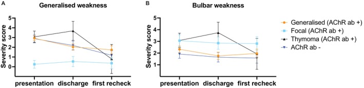
We classified 167 dogs into four subgroups: acetylcholine receptor (AChR) antibody-positive generalized (49.7%, n = 83/167), focal (19.2%, n = 32/167) and thymoma-associated MG (9%, n = 15/167) and seronegative MG (22.2%, n = 37/167). Dogs with thymoma-associated MG were older (median 102 months; Interquartile Range (IQR) 96-120; p < 0.001) and seronegative dogs were younger (median 30 months; IQR 11.5-66; p = 0.017), compared to the generalized subgroup (median 67 months; IQR 36-96). Seronegative dogs presented less frequently with megaesophagus, compared to the generalized subgroup (63.8% vs. 85.7%; Odds Ratio 3.4; 95% confidence intervals (C.I.) 1.4-8.9; p = 0.025). Myasthenic dogs' survival time was significantly reduced when thymoma (Hazard Ratio (H.R.) 3.7; 95% C.I. 1.4-9.9; p = 0.028) or esophageal weakness (H.R. 3.8; 95% C.I. 2.0-7.0; p < 0.001) was present. Conversely, a higher likelihood of remission was achieved when esophageal weakness was absent (H.R. 3.8; 95% C.I. 1.4-10.0; p = 0.007).
Dogs with seronegative MG are more common than previously reported. Myasthenic subgroups differ in presentation and outcome, with esophageal weakness key to survival and remission. Diagnostic tests for seronegative dogs and effective treatments for esophageal weakness in myasthenic dogs are urgently needed.
重症肌无力(MG)可分为几个亚组,包括血清阴性重症肌无力。血清阴性的人类患者已有充分记录,但血清阴性的犬类在临床上仍未得到充分描述,其患病率也未知。
本研究旨在评估犬重症肌无力亚组的临床表现、诊断、治疗及预后。
来自三个转诊中心的167只被诊断为重症肌无力的家养犬。
回顾性病例系列研究。我们按照人类指南将患重症肌无力的犬分为亚组。
我们将167只犬分为四个亚组:乙酰胆碱受体(AChR)抗体阳性全身型(49.7%,n = 83/167)、局灶型(19.2%,n = 32/167)、胸腺瘤相关型重症肌无力(9%,n = 15/167)和血清阴性重症肌无力(22.2%,n = 37/167)。与全身型亚组(中位年龄67个月;四分位间距(IQR)36 - 96)相比,胸腺瘤相关型重症肌无力的犬年龄更大(中位年龄102个月;IQR 96 - 120;p < 0.001),血清阴性的犬年龄更小(中位年龄30个月;IQR 11.5 - 66;p = 0.017)。与全身型亚组相比,血清阴性的犬发生巨食管的频率更低(63.8%对85.7%;优势比3.4;95%置信区间(C.I.)1.4 - 8.9;p = 0.025)。当存在胸腺瘤(风险比(H.R.)3.7;95% C.I. 1.4 - 9.9;p = 0.028)或食管肌无力(H.R. 3.8;95% C.I. 2.0 - 7.0;p < 0.001)时,患重症肌无力犬的生存时间显著缩短。相反,当不存在食管肌无力时,缓解的可能性更高(H.R. 3.8;95% C.I. 1.4 - 10.0;p = 0.007)。
血清阴性重症肌无力的犬比之前报道的更为常见。重症肌无力亚组在临床表现和预后方面存在差异,食管肌无力是生存和缓解的关键因素。迫切需要针对血清阴性犬的诊断测试以及针对患重症肌无力犬食管肌无力的有效治疗方法。