Kemmoku Emi, Kusumoto Shigeru, Kato Seiichi, Kawaguchi Yuka, Hagiwara Shinya, Saito Toko, Tokumasu Fukumi, Nonaka Ayako, Yanada Masamitsu, Kinoshita Tomohiro, Hosoda Waki, Yamamoto Kazuhito
Department of Hematology and Cell Therapy, Aichi Cancer Center Hospital, Nagoya, Japan.
Department of Pathology and Molecular Diagnostics, Aichi Cancer Center Hospital, Nagoya, Japan.
J Clin Exp Hematop. 2025 Jun 28;65(2):107-114. doi: 10.3960/jslrt.24065. Epub 2025 Apr 30.
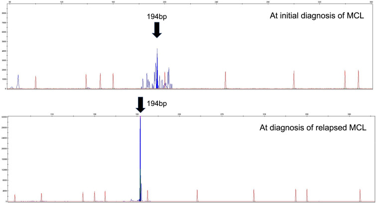
We encountered a patient with composite mantle cell lymphoma (MCL) and T-cell prolymphocytic leukemia (T-PLL) who presented with inactive disease to active T-PLL over 8 years. A 71-year-old man was diagnosed with MCL with an atypical T-cell population showing CD2+, CD3-, CD4+, CD7+, CD8-, and CD25+; however, the cause of the T-cell population could not be determined at the first MCL diagnosis. When MCL relapsed approximately 8 years after the initial treatment, T-PLL was definitively diagnosed using the T-PLL International Study Group criteria. MCL and T-PLL were determined to coexist in the lymph nodes and bone marrow by histological or flowcytometry analysis. Retrospective flow cytometry and T-cell receptor-polymerase chain reaction analysis of the stored samples suggested that the T-cell population noted at the time of initial MCL diagnosis eight years earlier was the same clone of T-PLL and the progression from inactive disease to active disease of his T-PLL. To the best of our knowledge, this is the first report of a composite MCL and T-PLL.
我们遇到了一位患有套细胞淋巴瘤(MCL)和T细胞幼淋巴细胞白血病(T-PLL)的患者,其疾病在8年多的时间里从静止期发展为活跃期的T-PLL。一名71岁男性被诊断为MCL,伴有非典型T细胞群,表现为CD2 +、CD3 -、CD4 +、CD7 +、CD8 -和CD25 +;然而,在首次诊断MCL时,无法确定T细胞群的原因。在初始治疗约8年后MCL复发时,根据T-PLL国际研究组标准明确诊断为T-PLL。通过组织学或流式细胞术分析确定MCL和T-PLL在淋巴结和骨髓中共存。对储存样本进行回顾性流式细胞术和T细胞受体聚合酶链反应分析表明,8年前首次诊断MCL时发现的T细胞群与T-PLL是同一克隆,且其T-PLL从静止期发展为活跃期。据我们所知,这是关于复合性MCL和T-PLL的首例报告。