Krishnan Aravind, Forouharshad Mahdi, Heng Elbert, Garrison Alyssa, Alnasir Daniel, Patil Shubham, Farazdaghi Arman, Fawad Moeed, Elde Stefan, Guenthart Brandon A, Ensign Laura M, Woo Y Joseph, Parikh Kunal S, MacArthur John W
Department of Cardiothoracic Surgery, Stanford University School of Medicine, Stanford, Calif.
Center for Nanomedicine at the Wilmer Eye Institute, Department of Ophthalmology, Johns Hopkins University School of Medicine, Baltimore, Md.
JTCVS Open. 2025 Jan 20;24:510-520. doi: 10.1016/j.xjon.2025.01.008. eCollection 2025 Apr.
Recipients of lung transplants experience the lowest long-term survival among all solid-organ transplant recipients. Airway complications contribute significantly to morbidity and mortality post-lung transplant and may be driven by airway devascularization inherent to procurement and implantation of the lungs. We studied application of biodegradable, nanofiber-based thin films to devascularized autotransplanted airways to mitigate airway ischemia.
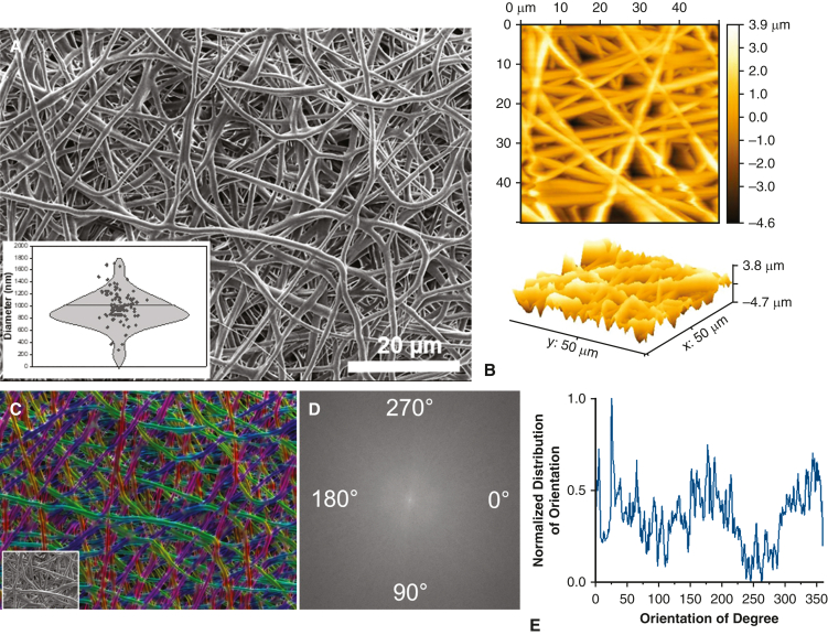
We used a rat tracheal autotransplantation model that replicates airway ischemia. Rats were divided into an operated control group (n = 18) and a treatment group (n = 12) receiving an electrospun film composed of randomly aligned polydioxanone (PDO) nanofibers applied to the circumferential surface of the transplanted trachea. Airway perfusion was assessed via laser speckle contrast analysis at 0, 3, and 10 days. Differences in perfusion units were calculated between the nontransplanted and transplanted segments of the trachea. Multimodal analysis of angiogenesis in tracheal autografts included immunoassay profiling for proangiogenic cytokines, histologic injury grading, and speckle angiography.
Qualitative and quantitative perfusion differences were demonstrated at days 0, 3, and 10. Nanofiber-based, PDO thin films significantly improved perfusion in the transplanted segment of trachea ( < .05). Histologic injury scoring was significantly worse in the operated controls compared with the treatment group ( < .01). Immunoassays demonstrated increased expression of vascular cell adhesion molecule 1 in the treatment group ( < .05).
Application of a nanofiber-based, PDO thin film induced a local tissue response that improved perfusion and histologic injury scoring of the transplanted airway in an autotransplant model of airway devascularization. Immune multiplexing suggests local inflammatory responses may drive angiogenesis.
在所有实体器官移植受者中,肺移植受者的长期生存率最低。气道并发症是肺移植后发病和死亡的重要原因,可能是由肺获取和植入过程中固有的气道血管离断所导致。我们研究了可生物降解的、基于纳米纤维的薄膜在自体移植离断气道中的应用,以减轻气道缺血。
我们使用了一种复制气道缺血的大鼠气管自体移植模型。将大鼠分为手术对照组(n = 18)和治疗组(n = 12),治疗组接受由随机排列的聚二氧六环酮(PDO)纳米纤维组成的电纺薄膜,该薄膜应用于移植气管的圆周表面。在第0、3和10天通过激光散斑对比分析评估气道灌注。计算气管未移植段和移植段之间灌注单位的差异。气管自体移植物血管生成的多模态分析包括促血管生成细胞因子的免疫测定分析、组织学损伤分级和散斑血管造影。
在第0、3和10天观察到定性和定量的灌注差异。基于纳米纤维的PDO薄膜显著改善了气管移植段的灌注(P <.05)。与治疗组相比,手术对照组的组织学损伤评分明显更差(P <.01)。免疫测定显示治疗组中血管细胞粘附分子1的表达增加(P <.05)。
在气道血管离断的自体移植模型中,应用基于纳米纤维的PDO薄膜可诱导局部组织反应,改善移植气道的灌注和组织学损伤评分。免疫多重分析表明局部炎症反应可能驱动血管生成。