Sakamoto Taku, Akiyama Shintaro, Narasaka Toshiaki, Tuchiya Kiichiro
Division of Gastroenterology University of Tsukuba Hospital Ibaraki Japan.
DEN Open. 2025 May 8;6(1):e70141. doi: 10.1002/deo2.70141. eCollection 2026 Apr.
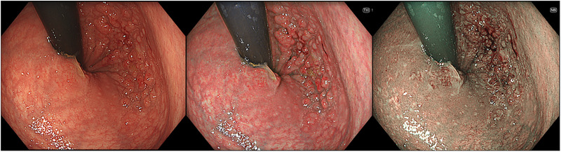
Colorectal cancer (CRC) is a leading cause of cancer-related mortality, highlighting the need for early detection and accurate lesion characterization. Traditional white-light imaging has limitations in detecting lesions, particularly those with flat morphology or minimal color contrast with the surrounding mucosa. It also struggles to distinguish neoplastic from non-neoplastic lesions. These limitations led to the development of image-enhanced endoscopy (IEE). Image-enhanced endoscopy modalities such as narrow-band imaging, blue laser imaging, linked color imaging, and texture and color enhancement imaging enhance mucosal surface and vascular pattern visualization, thereby improving lesion detection and characterization. In contrast, red dichromatic imaging is primarily designed to enhance the visibility of deep blood vessels, making it particularly useful during therapeutic endoscopies, such as identifying bleeding sources and monitoring post-treatment hemostasis. Although IEE enhances lesion detection and characterization, it remains limited in assessing submucosal invasion depth, which is a key factor in treatment decisions. Endoscopic submucosal dissection requires accurate prediction of invasion depth; however, IEE mainly reflects superficial features. Endoscopic ultrasound and artificial intelligence-assisted diagnostics have emerged as complementary techniques for improving depth assessment and lesion classification. Additionally, IEE plays a critical role in detecting ulcerative colitis-associated neoplasia (UCAN), which often presents with a flat morphology and indistinct borders. High-definition chromoendoscopy and IEE modalities enhance detection; however, inflammation-related changes limit diagnostic accuracy. Artificial intelligence and molecular biomarkers may improve UCAN diagnosis. This review examines the role of IEE in lesion detection and treatment selection, its limitations, and complementary techniques such as endoscopic ultrasound and artificial intelligence. We also explored pit pattern diagnosis using crystal violet staining and discussed emerging strategies to refine colorectal cancer screening and management.
结直肠癌(CRC)是癌症相关死亡的主要原因之一,这凸显了早期检测和准确病变特征描述的必要性。传统白光成像在检测病变方面存在局限性,尤其是那些形态扁平或与周围黏膜颜色对比度极小的病变。它也难以区分肿瘤性病变和非肿瘤性病变。这些局限性促使了图像增强内镜检查(IEE)的发展。窄带成像、蓝光成像、联动成像以及纹理和颜色增强成像等图像增强内镜检查方式可增强黏膜表面和血管形态的可视化,从而改善病变的检测和特征描述。相比之下,红色双色成像主要用于增强深部血管的可视性,在治疗性内镜检查中尤其有用,例如识别出血源和监测治疗后的止血情况。尽管IEE可增强病变的检测和特征描述,但在评估黏膜下浸润深度方面仍存在局限性,而黏膜下浸润深度是治疗决策的关键因素。内镜黏膜下剥离术需要准确预测浸润深度;然而,IEE主要反映的是表面特征。内镜超声和人工智能辅助诊断已成为改善深度评估和病变分类的补充技术。此外,IEE在检测溃疡性结肠炎相关肿瘤(UCAN)方面发挥着关键作用,UCAN通常形态扁平且边界不清晰。高清色素内镜检查和IEE方式可提高检测率;然而,炎症相关变化限制了诊断准确性。人工智能和分子生物标志物可能会改善UCAN的诊断。本综述探讨了IEE在病变检测和治疗选择中的作用、其局限性以及内镜超声和人工智能等补充技术。我们还探讨了使用结晶紫染色进行凹陷模式诊断,并讨论了完善结直肠癌筛查和管理的新兴策略。