• 文献检索
  • 文档翻译
  • 深度研究
  • 学术资讯
  • Suppr Zotero 插件Zotero 插件
  • 邀请有礼
  • 套餐&价格
  • 历史记录
应用&插件
Suppr Zotero 插件Zotero 插件浏览器插件Mac 客户端Windows 客户端微信小程序
定价
高级版会员购买积分包购买API积分包
服务
文献检索文档翻译深度研究API 文档MCP 服务
关于我们
关于 Suppr公司介绍联系我们用户协议隐私条款
关注我们

Suppr 超能文献

核心技术专利:CN118964589B侵权必究
粤ICP备2023148730 号-1Suppr @ 2026

文献检索

告别复杂PubMed语法,用中文像聊天一样搜索,搜遍4000万医学文献。AI智能推荐,让科研检索更轻松。

立即免费搜索

文件翻译

保留排版,准确专业,支持PDF/Word/PPT等文件格式,支持 12+语言互译。

免费翻译文档

深度研究

AI帮你快速写综述,25分钟生成高质量综述,智能提取关键信息,辅助科研写作。

立即免费体验

单细胞RNA测序和单细胞染色质可及性测序表明,在非梗阻性无精子症中,支持细胞通过阶段特异性通讯介导精子发生障碍。

scRNA-seq and scATAC-seq reveal that Sertoli cell mediates spermatogenesis disorders through stage-specific communications in non-obstructive azoospermia.

作者信息

Wang Shimin, Wang Hongxian, Jin Bicheng, Yan Hongli, Zheng Qingliang, Zhao Dong

机构信息

Prenatal Diagnosis Center, The Eighth Affiliated Hospital, Sun Yat-sen University, Shenzhen, China.

Department of Gynaecology and Obstetrics, Ninth People's Hospital, Shanghai Jiao Tong University School of Medicine, Shanghai, China.

出版信息

Elife. 2025 May 15;13:RP97958. doi: 10.7554/eLife.97958.

DOI:10.7554/eLife.97958
PMID:40371706
原文链接:https://pmc.ncbi.nlm.nih.gov/articles/PMC12081002/
Abstract

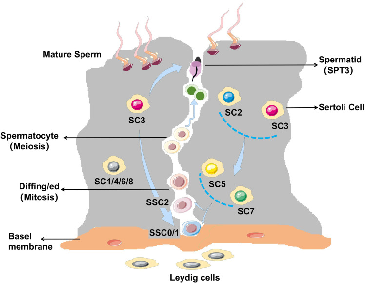
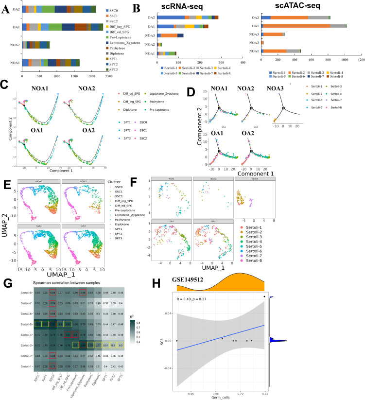
Non-obstructive azoospermia (NOA) belongs to male infertility due to spermatogenesis failure. However, evidence for cell type-specific abnormalities of spermatogenesis disorders in NOA remains lacking. We performed single-cell RNA sequencing (scRNA-seq) and single-cell assay for transposase-accessible chromatin sequencing (scATAC-seq) on testicular tissues from patients with obstructive azoospermia (OA) and NOA. HE staining confirmed the structural abnormalities of the seminiferous tubules in NOA patients. We identified 12 germ cell subtypes (spermatogonial stem cell-0 [SSC0], SSC1, SSC2, diffing-spermatogonia [Diffing-SPG], diffed-spermatogonia [Diffed-SPG], pre-leptotene [Pre-Lep], leptotene-zygotene [L-Z], pachytene [Pa], diplotene [Di], spermatids-1 [SPT1], SPT2, and SPT3) and 8 Sertoli cell subtypes (SC1-SC8). Among them, three novel Sertoli cell subtype phenotypes were identified, namely SC4/immature, SC7/mature, and SC8/further mature Sertoli cells. For each germ or Sertoli cell subtype, we identified unique new markers, among which immunofluorescence confirmed co-localization of ST3GAL4, A2M, ASB9, and TEX19 and DDX4 (classical marker of germ cell). PRAP1, BST2, and CCDC62 were co-expressed with SOX9 (classical marker of Sertoli cell) in testes tissues also confirmed by immunofluorescence. The interaction between germ cell subtypes and Sertoli cell subtypes exhibits stage-specific-matching pattern, as evidenced by SC1/2/5/7 involving in SSC0-2 development, SC3 participating in the whole process of spermiogenesis, SC4/6 participating in Diffing and Diffed-SPG development, and SC8 involving in the final stage of SPT3. This pattern of specific interactions between subtypes of germ cell and Sertoli cell was confirmed by immunofluorescence of novel markers in testes tissues. The interaction was mainly regulated by the Notch1/2/3 signaling. Our study profiled the single-cell transcriptome of human spermatogenesis and provided many potential molecular markers for developing testicular puncture-specific marker kits for NOA patients.

摘要

非梗阻性无精子症(NOA)属于因精子发生失败导致的男性不育症。然而,关于NOA中精子发生障碍的细胞类型特异性异常的证据仍然缺乏。我们对梗阻性无精子症(OA)和NOA患者的睾丸组织进行了单细胞RNA测序(scRNA-seq)和转座酶可及染色质测序的单细胞分析(scATAC-seq)。苏木精-伊红(HE)染色证实了NOA患者曲细精管的结构异常。我们鉴定出12种生殖细胞亚型(精原干细胞0 [SSC0]、SSC1、SSC2、分化型精原细胞[Diffing-SPG]、已分化精原细胞[Diffed-SPG]、前细线期[Pre-Lep]、细线期-偶线期[L-Z]、粗线期[Pa]、双线期[Di]、精子细胞1 [SPT1]、SPT2和SPT3)和8种支持细胞亚型(SC1-SC8)。其中,鉴定出三种新的支持细胞亚型表型,即SC4/未成熟型、SC7/成熟型和SC8/进一步成熟型支持细胞。对于每种生殖细胞或支持细胞亚型,我们鉴定出了独特的新标志物,其中免疫荧光证实了ST3GAL4、A2M、ASB9和TEX19与DDX4(生殖细胞的经典标志物)共定位。PRAP1、BST2和CCDC62也与SOX9(支持细胞的经典标志物)在睾丸组织中共表达,这也通过免疫荧光得到了证实。生殖细胞亚型与支持细胞亚型之间的相互作用呈现出阶段特异性匹配模式,如SC1/2/5/7参与SSC0-2的发育,SC3参与精子形成的全过程,SC4/6参与Diffing和Diffed-SPG的发育,SC8参与SPT3的最后阶段。生殖细胞和支持细胞亚型之间这种特异性相互作用模式通过睾丸组织中新标志物的免疫荧光得到了证实。这种相互作用主要受Notch1/2/3信号通路调控。我们的研究描绘了人类精子发生的单细胞转录组,并为开发针对NOA患者的睾丸穿刺特异性标志物试剂盒提供了许多潜在的分子标志物。

https://cdn.ncbi.nlm.nih.gov/pmc/blobs/188b/12081002/489504cca574/elife-97958-fig8.jpg
https://cdn.ncbi.nlm.nih.gov/pmc/blobs/188b/12081002/2f37c6127b9d/elife-97958-fig1.jpg
https://cdn.ncbi.nlm.nih.gov/pmc/blobs/188b/12081002/c01726dd6508/elife-97958-fig1-figsupp1.jpg
https://cdn.ncbi.nlm.nih.gov/pmc/blobs/188b/12081002/500db4c00abe/elife-97958-fig1-figsupp2.jpg
https://cdn.ncbi.nlm.nih.gov/pmc/blobs/188b/12081002/0459f3823810/elife-97958-fig2.jpg
https://cdn.ncbi.nlm.nih.gov/pmc/blobs/188b/12081002/5944443650fe/elife-97958-fig2-figsupp1.jpg
https://cdn.ncbi.nlm.nih.gov/pmc/blobs/188b/12081002/6defdf6cc6a8/elife-97958-fig2-figsupp2.jpg
https://cdn.ncbi.nlm.nih.gov/pmc/blobs/188b/12081002/98142c3985cc/elife-97958-fig2-figsupp3.jpg
https://cdn.ncbi.nlm.nih.gov/pmc/blobs/188b/12081002/1502a28894e0/elife-97958-fig2-figsupp4.jpg
https://cdn.ncbi.nlm.nih.gov/pmc/blobs/188b/12081002/0602e0e65154/elife-97958-fig2-figsupp5.jpg
https://cdn.ncbi.nlm.nih.gov/pmc/blobs/188b/12081002/ca3e59070b6a/elife-97958-fig3.jpg
https://cdn.ncbi.nlm.nih.gov/pmc/blobs/188b/12081002/4a60f49999e8/elife-97958-fig3-figsupp1.jpg
https://cdn.ncbi.nlm.nih.gov/pmc/blobs/188b/12081002/93651191b83a/elife-97958-fig4.jpg
https://cdn.ncbi.nlm.nih.gov/pmc/blobs/188b/12081002/93f1e437c540/elife-97958-fig4-figsupp1.jpg
https://cdn.ncbi.nlm.nih.gov/pmc/blobs/188b/12081002/ba377eec9814/elife-97958-fig4-figsupp2.jpg
https://cdn.ncbi.nlm.nih.gov/pmc/blobs/188b/12081002/27d3ae47aa77/elife-97958-fig4-figsupp3.jpg
https://cdn.ncbi.nlm.nih.gov/pmc/blobs/188b/12081002/6abf0cd2e28a/elife-97958-fig5.jpg
https://cdn.ncbi.nlm.nih.gov/pmc/blobs/188b/12081002/3310a0f93048/elife-97958-fig5-figsupp1.jpg
https://cdn.ncbi.nlm.nih.gov/pmc/blobs/188b/12081002/58932e0c2020/elife-97958-fig6.jpg
https://cdn.ncbi.nlm.nih.gov/pmc/blobs/188b/12081002/02f88620bbf1/elife-97958-fig6-figsupp1.jpg
https://cdn.ncbi.nlm.nih.gov/pmc/blobs/188b/12081002/d0513129aad3/elife-97958-fig6-figsupp2.jpg
https://cdn.ncbi.nlm.nih.gov/pmc/blobs/188b/12081002/03bffa51367c/elife-97958-fig6-figsupp3.jpg
https://cdn.ncbi.nlm.nih.gov/pmc/blobs/188b/12081002/48c374b8683e/elife-97958-fig6-figsupp4.jpg
https://cdn.ncbi.nlm.nih.gov/pmc/blobs/188b/12081002/d7b938fed455/elife-97958-fig6-figsupp5.jpg
https://cdn.ncbi.nlm.nih.gov/pmc/blobs/188b/12081002/16605b9c5657/elife-97958-fig7.jpg
https://cdn.ncbi.nlm.nih.gov/pmc/blobs/188b/12081002/489504cca574/elife-97958-fig8.jpg

相似文献

1
scRNA-seq and scATAC-seq reveal that Sertoli cell mediates spermatogenesis disorders through stage-specific communications in non-obstructive azoospermia.单细胞RNA测序和单细胞染色质可及性测序表明,在非梗阻性无精子症中,支持细胞通过阶段特异性通讯介导精子发生障碍。
Elife. 2025 May 15;13:RP97958. doi: 10.7554/eLife.97958.
2
Long-read single-cell sequencing reveals the transcriptional landscape of spermatogenesis in obstructive azoospermia and Sertoli cell-only patients.长读单细胞测序揭示梗阻性无精子症和唯支持细胞综合征患者精子发生的转录图谱。
QJM. 2024 Jun 25;117(6):422-435. doi: 10.1093/qjmed/hcae009.
3
Sertoli and Germ Cells Within Atrophic Seminiferous Tubules of Men With Non-Obstructive Azoospermia.非梗阻性无精子症患者曲细精管萎缩时的支持细胞和生殖细胞。
Front Endocrinol (Lausanne). 2022 Jun 2;13:825904. doi: 10.3389/fendo.2022.825904. eCollection 2022.
4
Transcriptome Analysis to Identify Human Spermatogonial Cells from Sertoli Cell-Only Testes.转录组分析鉴定支持细胞中仅有的人类精原细胞。
J Urol. 2020 Apr;203(4):809-816. doi: 10.1097/JU.0000000000000682. Epub 2019 Nov 19.
5
scRNA-Seq-Based Transcriptome Profiling and Relevant Bioinformatics Approaches to Uncover Novel Insights in Studying Human Spermatogenesis.基于单细胞RNA测序的转录组分析及相关生物信息学方法,以揭示人类精子发生研究中的新见解。
Adv Exp Med Biol. 2025;1469:173-205. doi: 10.1007/978-3-031-82990-1_9.
6
Investigation of CD133 and CD24 as candidate azoospermia markers and their relationship with spermatogenesis defects.探讨 CD133 和 CD24 作为非梗阻性无精子症候选标志物及其与精子发生缺陷的关系。
Gene. 2019 Jul 20;706:211-221. doi: 10.1016/j.gene.2019.04.028. Epub 2019 May 1.
7
Single-cell multi-omics analysis reveals dysfunctional Wnt signaling of spermatogonia in non-obstructive azoospermia.单细胞多组学分析揭示非梗阻性无精子症精原细胞中 Wnt 信号传导功能障碍。
Front Endocrinol (Lausanne). 2023 Jun 6;14:1138386. doi: 10.3389/fendo.2023.1138386. eCollection 2023.
8
Meiotic transcriptional reprogramming mediated by cell-cell communications in humans and mice revealed by scATAC-seq and scRNA-seq.通过 scATAC-seq 和 scRNA-seq 揭示人类和小鼠细胞间通讯介导的减数分裂转录重编程。
Zool Res. 2024 May 18;45(3):601-616. doi: 10.24272/j.issn.2095-8137.2023.414.
9
Evaluation of immune status in testis and macrophage polarization associated with testicular damage in patients with nonobstructive azoospermia.评价非梗阻性无精子症患者睾丸损伤相关的睾丸免疫状态和巨噬细胞极化。
Am J Reprod Immunol. 2021 Nov;86(5):e13481. doi: 10.1111/aji.13481. Epub 2021 Aug 9.
10
Single-cell transcriptome analysis of the novel coronavirus (SARS-CoV-2) associated gene ACE2 expression in normal and non-obstructive azoospermia (NOA) human male testes.新型冠状病毒(SARS-CoV-2)相关基因 ACE2 在正常和非梗阻性无精子症(NOA)男性睾丸中的单细胞转录组分析。
Sci China Life Sci. 2020 Jul;63(7):1006-1015. doi: 10.1007/s11427-020-1705-0. Epub 2020 Apr 30.

本文引用的文献

1
Understanding testicular single cell transcriptional atlas: from developmental complications to male infertility.理解睾丸单细胞转录图谱:从发育并发症到男性不育。
Front Endocrinol (Lausanne). 2024 Jul 11;15:1394812. doi: 10.3389/fendo.2024.1394812. eCollection 2024.
2
Preliminary experience with 3D digital image microscope system on the treatment of varicocele.3D数字图像显微镜系统治疗精索静脉曲张的初步经验。
Andrologia. 2022 Aug;54(7):1592-1597. doi: 10.1111/and.14423. Epub 2022 Mar 21.
3
Cytotoxicity of mancozeb on Sertoli-germ cell co-culture system: Role of MAPK signaling pathway.
代森锰锌对支持细胞-生殖细胞共培养体系的细胞毒性:MAPK 信号通路的作用。
Toxicol Ind Health. 2021 Nov;37(11):674-684. doi: 10.1177/07482337211044028. Epub 2021 Oct 13.
4
Sertoli cells as key drivers of testis function.支持细胞作为睾丸功能的关键驱动因素。
Semin Cell Dev Biol. 2022 Jan;121:2-9. doi: 10.1016/j.semcdb.2021.06.016. Epub 2021 Jul 3.
5
Scalable, multimodal profiling of chromatin accessibility, gene expression and protein levels in single cells.可扩展的、多模式的单细胞染色质可及性、基因表达和蛋白水平的分析。
Nat Biotechnol. 2021 Oct;39(10):1246-1258. doi: 10.1038/s41587-021-00927-2. Epub 2021 Jun 3.
6
Omics in Seminal Plasma: An Effective Strategy for Predicting Sperm Retrieval Outcome in Non-obstructive Azoospermia.精液血浆组学:预测非梗阻性无精子症精子获取结果的有效策略。
Mol Diagn Ther. 2021 May;25(3):315-325. doi: 10.1007/s40291-021-00524-8. Epub 2021 Apr 15.
7
An Insight into the Role of UTF1 in Development, Stem Cells, and Cancer.深入探讨 UTF1 在发育、干细胞和癌症中的作用。
Stem Cell Rev Rep. 2021 Aug;17(4):1280-1293. doi: 10.1007/s12015-021-10127-9. Epub 2021 Jan 30.
8
Single-cell analysis of the developing human testis reveals somatic niche cell specification and fetal germline stem cell establishment.单细胞分析人类睾丸的发育过程揭示了体细胞龛细胞的特化和胎儿生殖细胞干细胞的建立。
Cell Stem Cell. 2021 Apr 1;28(4):764-778.e4. doi: 10.1016/j.stem.2020.12.004. Epub 2021 Jan 15.
9
Single-cell analysis of developing and azoospermia human testicles reveals central role of Sertoli cells.单细胞分析发育和无精子症人类睾丸揭示了支持细胞的核心作用。
Nat Commun. 2020 Nov 10;11(1):5683. doi: 10.1038/s41467-020-19414-4.
10
Integrated Single-Cell Transcriptomics and Chromatin Accessibility Analysis Reveals Regulators of Mammary Epithelial Cell Identity.单细胞转录组学和染色质可及性分析的整合揭示了乳腺上皮细胞身份的调控因子。
Cell Rep. 2020 Oct 20;33(3):108273. doi: 10.1016/j.celrep.2020.108273.