• 文献检索
  • 文档翻译
  • 深度研究
  • 学术资讯
  • Suppr Zotero 插件Zotero 插件
  • 邀请有礼
  • 套餐&价格
  • 历史记录
应用&插件
Suppr Zotero 插件Zotero 插件浏览器插件Mac 客户端Windows 客户端微信小程序
定价
高级版会员购买积分包购买API积分包
服务
文献检索文档翻译深度研究API 文档MCP 服务
关于我们
关于 Suppr公司介绍联系我们用户协议隐私条款
关注我们

Suppr 超能文献

核心技术专利:CN118964589B侵权必究
粤ICP备2023148730 号-1Suppr @ 2026

文献检索

告别复杂PubMed语法,用中文像聊天一样搜索,搜遍4000万医学文献。AI智能推荐,让科研检索更轻松。

立即免费搜索

文件翻译

保留排版,准确专业,支持PDF/Word/PPT等文件格式,支持 12+语言互译。

免费翻译文档

深度研究

AI帮你快速写综述,25分钟生成高质量综述,智能提取关键信息,辅助科研写作。

立即免费体验

阻塞性睡眠呼吸暂停患者皮质下灰质核团变化的合成磁共振成像初步研究。

A preliminary study of synthetic magnetic resonance imaging on the changes of subcortical gray matter nuclei in obstructive sleep apnea patients.

作者信息

Peng Kun, Zhang Ning, Liu Qing, Xiao Ailian, Wang Kaiyu, Zhang Hui

机构信息

College of Medical Imaging, Shanxi Medical University, Taiyuan, China.

Department of Radiology, The Sixth Hospital of Shanxi Medical University (General Hospital of TISCO), Taiyuan, China.

出版信息

Quant Imaging Med Surg. 2025 May 1;15(5):3911-3922. doi: 10.21037/qims-24-1830. Epub 2025 Apr 14.

DOI:10.21037/qims-24-1830
PMID:40384657
原文链接:https://pmc.ncbi.nlm.nih.gov/articles/PMC12082584/
Abstract

BACKGROUND

Obstructive sleep apnea (OSA) is a relatively common sleep disorder, which can damage the brain structure and thus trigger cognitive impairment. We aimed to assess changes in volume and relaxation values of subcortical gray matter nuclei in patients with moderate to severe OSA using synthetic magnetic resonance imaging (SyMRI).

METHODS

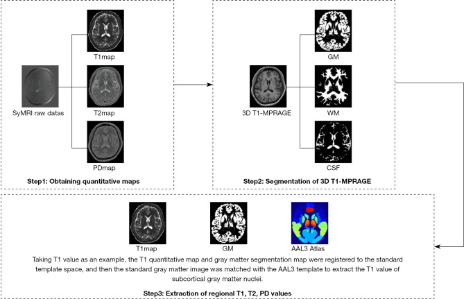
A total of 30 patients with moderate to severe OSA, newly diagnosed by polysomnography (PSG), and 30 age-, sex-, education-, and handedness-matched healthy controls (HC) were recruited. All participants underwent the Montreal Cognitive Assessment (MoCA) Scale. Conventional MRI, three-dimensional T1-weighted brain volume (3D T1-BRAVO), and SyMRI were performed on both groups using a 3.0TMR scanner. After scanning, original SyMRI data were post-processed using SyMRI8.0 software to automatically generate T1, T2, and proton density (PD) quantitative maps, and subcortical gray matter nuclei were obtained using SPM12 software (Matlab R2015b). Cognitive scale scores, subcortical gray matter nuclei volume, and relaxation quantitative values were compared between groups. Volume and relaxation quantitative values were corrected for multiple comparisons using a false discovery rate (FDR). SyMRI quantitative parameters with statistically significant differences underwent receiver operating characteristic (ROC) analysis to calculate the area under the curve (AUC). Correlations between changes in abnormal brain volume and relaxation values and MoCA scores were analyzed in the OSA group, using FDR for multiple comparisons and corrections.

RESULTS

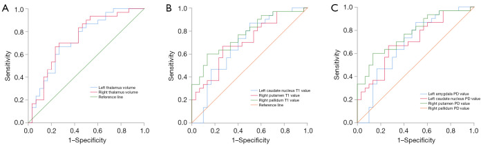
Compared to controls, the OSA group exhibited a significant decrease in bilateral thalamic volume (P=0.002, 0.003, uncorrected). T1 values increased significantly in the left hippocampus, amygdala, caudate nucleus, right pallidum, thalamus, and bilateral putamen (P=0.02, 0.040, 0.01, <0.001, 0.03, 0.04, 0.004, uncorrected). The left hippocampus showed significantly increased T2 values (P=0.03, uncorrected). In contrast, the PD values increased significantly in the left amygdala, nucleus accumbens, right pallidum, putamen, bilateral caudate nucleus, and thalamus (P=0.02, 0.041, 0.01, 0.006, 0.007, 0.03, 0.047, 0.009, uncorrected). After FDR correction, significant differences persisted in the bilateral thalamic volume, T1 values of left caudate, right putamen and pallidum, PD values of left amygdala, right pallidum, putamen, bilateral caudate nucleus, and thalamus. ROC curve analysis revealed significant differences in bilateral thalamic volume, T1 values of left caudate nucleus, right putamen and pallidum, PD values of left amygdala, caudate nucleus, right putamen, pallidum, and thalamus between OSA patients and controls (P=0.002, 0.001, 0.01, 0.007, <0.001, 0.02, 0.007, 0.01, 0.01, 0.03; AUC 0.668-0.770). After controlling for age, body mass index (BMI), and years of education, OSA patients showed negative correlations between visual space and executive function and the right putamen T1 and PD values (r=-0.390, -0.449; P=0.045, 0.02) and positive correlations with the left amygdala PD value (r=0.397; P=0.04). No significant differences were found in partial correlation analysis after FDR correction.

CONCLUSIONS

SyMRI offers sensitive detection of abnormal volume and relaxation value changes in subcortical gray matter nuclei among patients with moderate to severe OSA. These findings provide valuable imaging information for quantifying subcortical gray matter nuclei damage in OSA and advancing our understanding of the neuropathological mechanisms underlying cognitive impairment.

https://cdn.ncbi.nlm.nih.gov/pmc/blobs/285e/12082584/d82b0107e78b/qims-15-05-3911-f3.jpg
https://cdn.ncbi.nlm.nih.gov/pmc/blobs/285e/12082584/b70c20220984/qims-15-05-3911-f1.jpg
https://cdn.ncbi.nlm.nih.gov/pmc/blobs/285e/12082584/e9c3c979b9f6/qims-15-05-3911-f2.jpg
https://cdn.ncbi.nlm.nih.gov/pmc/blobs/285e/12082584/d82b0107e78b/qims-15-05-3911-f3.jpg
https://cdn.ncbi.nlm.nih.gov/pmc/blobs/285e/12082584/b70c20220984/qims-15-05-3911-f1.jpg
https://cdn.ncbi.nlm.nih.gov/pmc/blobs/285e/12082584/e9c3c979b9f6/qims-15-05-3911-f2.jpg
https://cdn.ncbi.nlm.nih.gov/pmc/blobs/285e/12082584/d82b0107e78b/qims-15-05-3911-f3.jpg
摘要

背景

阻塞性睡眠呼吸暂停(OSA)是一种相对常见的睡眠障碍,可损害脑结构并引发认知障碍。我们旨在使用合成磁共振成像(SyMRI)评估中度至重度OSA患者皮质下灰质核团的体积和弛豫值变化。

方法

招募了30例经多导睡眠监测(PSG)新诊断的中度至重度OSA患者,以及30例年龄、性别、教育程度和利手匹配的健康对照(HC)。所有参与者均接受蒙特利尔认知评估(MoCA)量表测试。两组均使用3.0T磁共振扫描仪进行常规MRI、三维T1加权脑容积(3D T1-BRAVO)和SyMRI检查。扫描后,使用SyMRI8.0软件对原始SyMRI数据进行后处理,以自动生成T1、T2和质子密度(PD)定量图,并使用SPM12软件(Matlab R2015b)获取皮质下灰质核团。比较两组之间的认知量表评分、皮质下灰质核团体积和弛豫定量值。使用错误发现率(FDR)对体积和弛豫定量值进行多重比较校正。对具有统计学显著差异的SyMRI定量参数进行受试者操作特征(ROC)分析,以计算曲线下面积(AUC)。在OSA组中,分析异常脑体积和弛豫值变化与MoCA评分之间的相关性,使用FDR进行多重比较和校正。

结果

与对照组相比,OSA组双侧丘脑体积显著减小(P=0.002,0.003,未校正)。左侧海马体、杏仁核、尾状核、右侧苍白球、丘脑以及双侧壳核的T1值显著升高(P=0.02,0.040,0.01,<0.001,0.03,0.04,0.004,未校正)。左侧海马体的T2值显著升高(P=0.03,未校正)。相比之下,左侧杏仁核、伏隔核、右侧苍白球、壳核、双侧尾状核和丘脑的PD值显著升高(P=0.02,0.041,0.01,0.006,0.007,0.03,0.047,0.009,未校正)。经FDR校正后,双侧丘脑体积、左侧尾状核、右侧壳核和苍白球的T1值、左侧杏仁核、右侧苍白球、壳核、双侧尾状核和丘脑的PD值仍存在显著差异。ROC曲线分析显示,OSA患者与对照组之间在双侧丘脑体积、左侧尾状核、右侧壳核和苍白球的T1值、左侧杏仁核、尾状核、右侧壳核、苍白球和丘脑的PD值方面存在显著差异(P=0.002,0.001,0.01,0.007,<0.001,0.02,0.007,0.01,0.01,0.03;AUC 0.668 - 0.770)。在控制年龄、体重指数(BMI)和教育年限后,OSA患者的视觉空间与执行功能与右侧壳核T1和PD值呈负相关(r=-0.390,-0.449;P=0.045,0.02),与左侧杏仁核PD值呈正相关(r=0.397;P=0.04)。FDR校正后的偏相关分析未发现显著差异。

结论

SyMRI能够灵敏检测中度至重度OSA患者皮质下灰质核团的异常体积和弛豫值变化。这些发现为量化OSA患者皮质下灰质核团损伤以及增进我们对认知障碍潜在神经病理机制的理解提供了有价值的影像学信息。

相似文献

1
A preliminary study of synthetic magnetic resonance imaging on the changes of subcortical gray matter nuclei in obstructive sleep apnea patients.阻塞性睡眠呼吸暂停患者皮质下灰质核团变化的合成磁共振成像初步研究。
Quant Imaging Med Surg. 2025 May 1;15(5):3911-3922. doi: 10.21037/qims-24-1830. Epub 2025 Apr 14.
2
Are there any differences at gray matter sites between severe obstructive sleep apnea patients and healthy controls?严重阻塞性睡眠呼吸暂停患者与健康对照者的灰质部位有差异吗?
Sleep Med. 2024 Apr;116:27-31. doi: 10.1016/j.sleep.2024.02.021. Epub 2024 Feb 17.
3
Subcortical nuclei in Alzheimer's disease: a volumetric and diffusion kurtosis imaging study.阿尔茨海默病中的皮质下核团:一项容积和扩散峰度成像研究。
Acta Radiol. 2018 Nov;59(11):1365-1371. doi: 10.1177/0284185118758122. Epub 2018 Feb 26.
4
[A 3.0 T MRI study on the alterations of the volume and morphology of fifteen subcortical nucleus in patients with early post-stroke depression].[一项关于脑卒中后早期抑郁患者15个皮质下核体积和形态改变的3.0T磁共振成像研究]
Zhonghua Yi Xue Za Zhi. 2018 Aug 21;98(31):2471-2475. doi: 10.3760/cma.j.issn.0376-2491.2018.31.004.
5
Diagnosis of Subcortical Ischemic Vascular Cognitive Impairment With No Dementia Using Radiomics of Cerebral Cortex and Subcortical Nuclei in High-Resolution T1-Weighted MR Imaging.利用高分辨率T1加权磁共振成像中大脑皮层和皮层下核团的影像组学诊断无痴呆的皮质下缺血性血管性认知障碍
Front Oncol. 2022 Apr 8;12:852726. doi: 10.3389/fonc.2022.852726. eCollection 2022.
6
Discriminative analysis of early-stage Alzheimer's disease and normal aging with automatic segmentation technique in subcortical gray matter structures: a multicenter in vivo MRI volumetric and DTI study.利用自动分割技术对早期阿尔茨海默病与正常衰老在皮质下灰质结构中的判别分析:一项多中心活体MRI容积和DTI研究
Acta Radiol. 2013 Dec;54(10):1191-200. doi: 10.1177/0284185113492971. Epub 2013 Jul 22.
7
Changes in the Shape and Volume of Subcortical Structures in Patients With End-Stage Renal Disease.终末期肾病患者皮质下结构的形状和体积变化
Front Hum Neurosci. 2021 Dec 16;15:778807. doi: 10.3389/fnhum.2021.778807. eCollection 2021.
8
Heritability of Subcortical Grey Matter Structures.脑区灰质结构的遗传度。
Medicina (Kaunas). 2022 Nov 21;58(11):1687. doi: 10.3390/medicina58111687.
9
Regional high iron deposition on quantitative susceptibility mapping correlates with cognitive decline in type 2 diabetes mellitus.定量磁化率成像上的局部高铁沉积与2型糖尿病患者的认知功能下降相关。
Front Neurosci. 2023 Jan 30;17:1061156. doi: 10.3389/fnins.2023.1061156. eCollection 2023.
10
Characterizing thalamic and basal ganglia nuclei in medically intractable focal epilepsy by MR fingerprinting.采用磁共振指纹技术对药物难治性局灶性癫痫进行丘脑和基底节核团特征分析。
Epilepsia. 2022 Aug;63(8):1998-2010. doi: 10.1111/epi.17318. Epub 2022 Jun 26.

本文引用的文献

1
Assessing Cognitive Impairments in Obstructive Sleep Apnea Patients Using Montreal Cognitive Assessment (MoCA) Scores.使用蒙特利尔认知评估量表(MoCA)评分评估阻塞性睡眠呼吸暂停患者的认知障碍
Cureus. 2024 Sep 24;16(9):e70085. doi: 10.7759/cureus.70085. eCollection 2024 Sep.
2
Obstructive sleep apnea and obesity: A review of epidemiology, pathophysiology and the effect of weight-loss treatments.阻塞性睡眠呼吸暂停与肥胖:流行病学、病理生理学及减肥治疗效果的综述。
Sleep Med Rev. 2024 Dec;78:101996. doi: 10.1016/j.smrv.2024.101996. Epub 2024 Aug 30.
3
Microstructural brain abnormalities and associated neurocognitive dysfunction in obstructive sleep apnea: a pilot study with diffusion kurtosis imaging.
阻塞性睡眠呼吸暂停患者的脑微观结构异常及相关神经认知功能障碍:一项基于扩散峰度成像的初步研究。
J Clin Sleep Med. 2024 Oct 1;20(10):1571-1578. doi: 10.5664/jcsm.11184.
4
Obstructive sleep apnea and dementia: A role to play?阻塞性睡眠呼吸暂停与痴呆:有一定作用?
Rev Neurol (Paris). 2023 Oct;179(7):793-803. doi: 10.1016/j.neurol.2023.08.006. Epub 2023 Aug 24.
5
Brain tissue integrity mapping in adults with obstructive sleep apnea using T1-weighted and T2-weighted images.利用T1加权和T2加权图像对患有阻塞性睡眠呼吸暂停的成年人进行脑组织完整性映射。
Ther Adv Neurol Disord. 2022 Nov 19;15:17562864221137505. doi: 10.1177/17562864221137505. eCollection 2022.
6
Screening for Obstructive Sleep Apnea in Adults: US Preventive Services Task Force Recommendation Statement.成人阻塞性睡眠呼吸暂停筛查:美国预防服务工作组推荐声明。
JAMA. 2022 Nov 15;328(19):1945-1950. doi: 10.1001/jama.2022.20304.
7
Alterations of Limbic Structure Volumes in Patients with Obstructive Sleep Apnea.阻塞性睡眠呼吸暂停患者边缘结构体积的改变。
Can J Neurol Sci. 2023 Sep;50(5):730-737. doi: 10.1017/cjn.2022.303. Epub 2022 Oct 17.
8
The Link between Obstructive Sleep Apnea and Neurocognitive Impairment: An Official American Thoracic Society Workshop Report.阻塞性睡眠呼吸暂停与神经认知障碍的关联:美国胸科学会官方专题研讨会报告。
Ann Am Thorac Soc. 2022 Aug;19(8):1245-1256. doi: 10.1513/AnnalsATS.202205-380ST.
9
Bidirectional relationships of comorbidity with obstructive sleep apnoea.共病与阻塞性睡眠呼吸暂停的双向关系。
Eur Respir Rev. 2022 May 4;31(164). doi: 10.1183/16000617.0256-2021. Print 2022 Jun 30.
10
Quantitative Analysis of Synthetic Magnetic Resonance Imaging in Alzheimer's Disease.阿尔茨海默病中合成磁共振成像的定量分析
Front Aging Neurosci. 2021 Apr 12;13:638731. doi: 10.3389/fnagi.2021.638731. eCollection 2021.