• 文献检索
  • 文档翻译
  • 深度研究
  • 学术资讯
  • Suppr Zotero 插件Zotero 插件
  • 邀请有礼
  • 套餐&价格
  • 历史记录
应用&插件
Suppr Zotero 插件Zotero 插件浏览器插件Mac 客户端Windows 客户端微信小程序
定价
高级版会员购买积分包购买API积分包
服务
文献检索文档翻译深度研究API 文档MCP 服务
关于我们
关于 Suppr公司介绍联系我们用户协议隐私条款
关注我们

Suppr 超能文献

核心技术专利:CN118964589B侵权必究
粤ICP备2023148730 号-1Suppr @ 2026

文献检索

告别复杂PubMed语法,用中文像聊天一样搜索,搜遍4000万医学文献。AI智能推荐,让科研检索更轻松。

立即免费搜索

文件翻译

保留排版,准确专业,支持PDF/Word/PPT等文件格式,支持 12+语言互译。

免费翻译文档

深度研究

AI帮你快速写综述,25分钟生成高质量综述,智能提取关键信息,辅助科研写作。

立即免费体验

基于常规超声和超声造影的甲状腺良恶性结节鉴别列线图的开发与验证

Development and validation of a nomogram based on conventional and contrast-enhanced ultrasound for differentiating malignant from benign thyroid nodules.

作者信息

Wang Qi-Guo, Li Mei, Deng Guang-Xiu, Huang Hai-Qing, Qiu Qin, Lin Jian-Jun

机构信息

Department of Medical Ultrasound, the First People's Hospital of Qinzhou, Qinzhou, China.

Department of Medical Ultrasound, the People's Hospital of Chongzuo, Chongzuo, China.

出版信息

Quant Imaging Med Surg. 2025 May 1;15(5):4641-4654. doi: 10.21037/qims-24-1796. Epub 2025 Apr 28.

DOI:10.21037/qims-24-1796
PMID:40384666
原文链接:https://pmc.ncbi.nlm.nih.gov/articles/PMC12082573/
Abstract

BACKGROUND

Conventional ultrasound (US) has been routinely used for differential diagnosis of thyroid nodules, but its discriminatory performance remains unsatisfactory. This study aimed to develop and validate a prediction nomogram model based on conventional US and contrast-enhanced ultrasound (CEUS) features for differentiating malignant from benign thyroid nodules.

METHODS

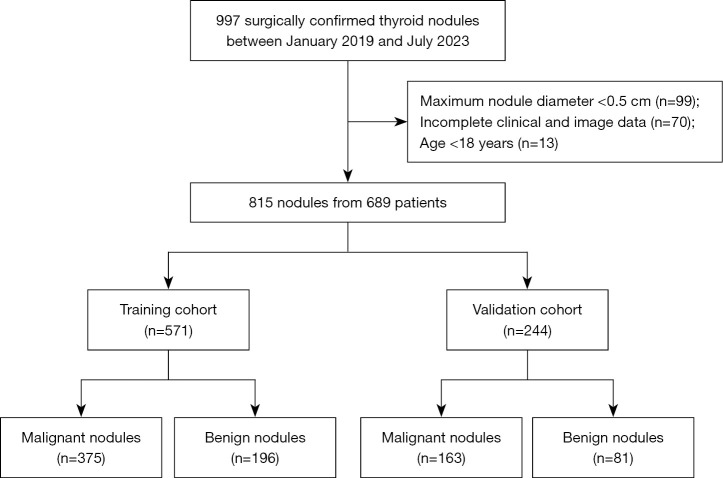
A total of 815 thyroid nodules with surgical pathology results and complete conventional US and CEUS data were retrospectively collected from the First People's Hospital of Qinzhou between January 2019 and July 2023. The nodules were grouped into a training cohort (n=571) and a validation cohort (n=244) at a 7:3 ratio. Independent risk factors of malignancy were selected by stepwise multivariate logistic regression analysis, and a prediction nomogram model was subsequently constructed. The diagnostic performance of the model was evaluated by the area under the receiver operating characteristic curve (AUC) in both the training and validation cohorts. The unnecessary fine-needle aspiration biopsy (FNAB) rate was calculated.

RESULTS

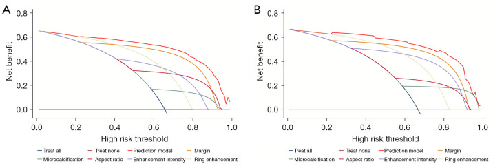
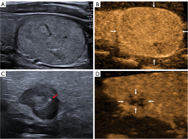
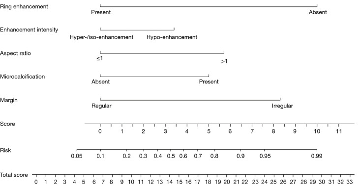
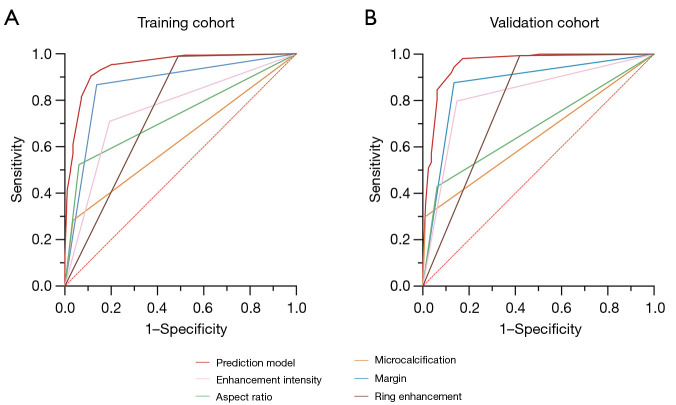
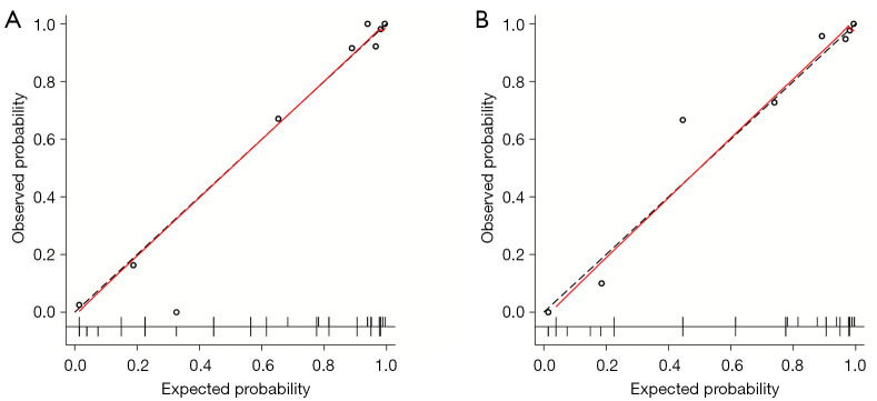
Multivariate logistic regression analysis identified irregular margin, aspect ratio >1, and microcalcification from conventional US images, as well as hypo-enhancement intensity and ring enhancement from CEUS images, as independent predictors for malignancy. The AUC, sensitivity, specificity, and accuracy of the prediction nomogram model were 0.947 [95% confidence interval (CI): 0.928-0.966], 90.4%, 88.8%, and 89.8% in the training cohort, and 0.957 (95% CI: 0.928-0.986), 94.5%, 86.4%, and 91.8% in the validation cohort, respectively. Using the prediction model, the unnecessary FNAB rates reduced from 29.6% to 6.1% in the training cohort and from 29.3% to 6.7% in the validation cohort compared to the Chinese Thyroid Imaging Reporting and Data System. Decision curve analysis demonstrated good clinical utility of the nomogram model.

CONCLUSIONS

The prediction nomogram model incorporating conventional US and CEUS features could effectively distinguish between malignant and benign thyroid nodules and reduce unnecessary FNAB rates.

摘要

背景

传统超声(US)一直被常规用于甲状腺结节的鉴别诊断,但其鉴别性能仍不尽人意。本研究旨在开发并验证一种基于传统超声和对比增强超声(CEUS)特征的预测列线图模型,用于区分甲状腺恶性结节和良性结节。

方法

回顾性收集了2019年1月至2023年7月期间钦州市第一人民医院的815个具有手术病理结果以及完整传统超声和CEUS数据的甲状腺结节。这些结节按7:3的比例分为训练队列(n = 571)和验证队列(n = 244)。通过逐步多因素逻辑回归分析选择恶性肿瘤的独立危险因素,随后构建预测列线图模型。在训练队列和验证队列中,通过受试者操作特征曲线(AUC)下面积评估模型的诊断性能。计算不必要的细针穿刺活检(FNAB)率。

结果

多因素逻辑回归分析确定,传统超声图像中的边界不规则、纵横比>1和微钙化,以及CEUS图像中的低增强强度和环状增强是恶性肿瘤的独立预测因素。预测列线图模型在训练队列中的AUC、敏感性、特异性和准确性分别为0.947 [95%置信区间(CI):0.928 - 0.966]、90.4%、88.8%和89.8%,在验证队列中分别为0.957(95% CI:0.928 - 0.986)、94.5%、86.4%和91.8%。与中国甲状腺影像报告和数据系统相比,使用预测模型后,训练队列中的不必要FNAB率从29.6%降至6.1%,验证队列中从29.3%降至6.7%。决策曲线分析表明列线图模型具有良好的临床实用性。

结论

结合传统超声和CEUS特征的预测列线图模型能够有效区分甲状腺恶性结节和良性结节,并降低不必要的FNAB率。

https://cdn.ncbi.nlm.nih.gov/pmc/blobs/dc51/12082573/85c0f16a0222/qims-15-05-4641-f6.jpg
https://cdn.ncbi.nlm.nih.gov/pmc/blobs/dc51/12082573/efeaf66427c7/qims-15-05-4641-f1.jpg
https://cdn.ncbi.nlm.nih.gov/pmc/blobs/dc51/12082573/860c4e24dddc/qims-15-05-4641-f2.jpg
https://cdn.ncbi.nlm.nih.gov/pmc/blobs/dc51/12082573/7373f37fa133/qims-15-05-4641-f3.jpg
https://cdn.ncbi.nlm.nih.gov/pmc/blobs/dc51/12082573/d5a47b50d940/qims-15-05-4641-f4.jpg
https://cdn.ncbi.nlm.nih.gov/pmc/blobs/dc51/12082573/0495cf6ecc84/qims-15-05-4641-f5.jpg
https://cdn.ncbi.nlm.nih.gov/pmc/blobs/dc51/12082573/85c0f16a0222/qims-15-05-4641-f6.jpg

相似文献

1
Development and validation of a nomogram based on conventional and contrast-enhanced ultrasound for differentiating malignant from benign thyroid nodules.基于常规超声和超声造影的甲状腺良恶性结节鉴别列线图的开发与验证
Quant Imaging Med Surg. 2025 May 1;15(5):4641-4654. doi: 10.21037/qims-24-1796. Epub 2025 Apr 28.
2
Development and Validation of a Nomogram Based on Multimodality Ultrasonography Images for Differentiating Malignant from Benign American College of Radiology Thyroid Imaging, Reporting and Data System (TI-RADS) 3-5 Thyroid Nodules.基于多模态超声图像鉴别良恶性美国放射学院甲状腺影像报告和数据系统(TI-RADS)3-5 级甲状腺结节的列线图的建立与验证。
Ultrasound Med Biol. 2024 Apr;50(4):557-563. doi: 10.1016/j.ultrasmedbio.2023.12.020. Epub 2024 Jan 22.
3
Dual-modal radiomics nomogram based on contrast-enhanced ultrasound to improve differential diagnostic accuracy and reduce unnecessary biopsy rate in ACR TI-RADS 4-5 thyroid nodules.基于超声造影的双模态影像组学列线图提高 ACR TI-RADS 4-5 甲状腺结节的鉴别诊断准确性并降低不必要的活检率
Cancer Imaging. 2024 Jan 23;24(1):17. doi: 10.1186/s40644-024-00661-3.
4
The diagnostic value of a nomogram based on multimodal ultrasonography for thyroid-nodule differentiation: A multicenter study.基于多模态超声的列线图对甲状腺结节鉴别诊断的价值:一项多中心研究
Front Oncol. 2022 Aug 18;12:970758. doi: 10.3389/fonc.2022.970758. eCollection 2022.
5
Predictors and a prediction model for positive fine needle aspiration biopsy in C-TIRADS 4 thyroid nodules.C-TIRADS 4 类甲状腺结节中细针抽吸活检阳性的预测因素及预测模型。
Front Endocrinol (Lausanne). 2023 Jul 24;14:1154984. doi: 10.3389/fendo.2023.1154984. eCollection 2023.
6
Contrast-Enhanced Ultrasound in the Differential Diagnosis and Risk Stratification of ACR TI-RADS Category 4 and 5 Thyroid Nodules With Non-Hypovascular.非低血供的ACR TI-RADS 4类和5类甲状腺结节鉴别诊断及风险分层中的超声造影
Front Oncol. 2021 May 26;11:662273. doi: 10.3389/fonc.2021.662273. eCollection 2021.
7
Diagnostic efficacy of the contrast-enhanced ultrasound thyroid imaging reporting and data system classification for benign and malignant thyroid nodules.超声造影甲状腺影像报告和数据系统分类对甲状腺良恶性结节的诊断效能
Quant Imaging Med Surg. 2024 Aug 1;14(8):5721-5736. doi: 10.21037/qims-24-457. Epub 2024 Jul 30.
8
Contrast-Enhanced Ultrasound for Diagnosing Thyroid Nodules With Indeterminate Cytology: A Retrospective Study.超声造影诊断甲状腺细针穿刺活检结果不确定的甲状腺结节:一项回顾性研究
Clin Endocrinol (Oxf). 2025 Feb;102(2):223-231. doi: 10.1111/cen.15160. Epub 2024 Nov 6.
9
Radiomics Combined with ACR TI-RADS for Thyroid Nodules: Diagnostic Performance, Unnecessary Biopsy Rate, and Nomogram Construction.基于影像组学与 ACR TI-RADS 联合应用对甲状腺结节的诊断效能、降低不必要的活检率及构建列线图的研究
Acad Radiol. 2024 Dec;31(12):4856-4865. doi: 10.1016/j.acra.2024.07.053. Epub 2024 Oct 4.
10
A Comparative Study of Two Radiomics-Based Blood Flow Modes with Thyroid Imaging Reporting and Data System in Predicting Malignancy of Thyroid Nodules and Reducing Unnecessary Fine-Needle Aspiration Rate.基于两种影像组学血流模式的对比研究与甲状腺影像报告和数据系统在预测甲状腺结节良恶性及降低不必要的细针抽吸率中的应用
Acad Radiol. 2024 Jul;31(7):2739-2752. doi: 10.1016/j.acra.2024.02.007. Epub 2024 Mar 7.

本文引用的文献

1
Comparison of diagnostic performance of the current score-based ultrasound risk stratification systems according to thyroid nodule size.根据甲状腺结节大小比较当前基于评分的超声风险分层系统的诊断性能。
Quant Imaging Med Surg. 2024 Dec 5;14(12):9234-9245. doi: 10.21037/qims-24-282. Epub 2024 Nov 6.
2
Diagnostic efficacy of the contrast-enhanced ultrasound thyroid imaging reporting and data system classification for benign and malignant thyroid nodules.超声造影甲状腺影像报告和数据系统分类对甲状腺良恶性结节的诊断效能
Quant Imaging Med Surg. 2024 Aug 1;14(8):5721-5736. doi: 10.21037/qims-24-457. Epub 2024 Jul 30.
3
Diagnostic value of contrast-enhanced ultrasound in the diagnosis of papillary thyroid microcarcinoma: A systematic review and meta-analysis.
超声造影对甲状腺微小乳头状癌的诊断价值:系统评价和荟萃分析。
Medicine (Baltimore). 2024 Apr 12;103(15):e37768. doi: 10.1097/MD.0000000000037768.
4
A Comparative Study of Two Radiomics-Based Blood Flow Modes with Thyroid Imaging Reporting and Data System in Predicting Malignancy of Thyroid Nodules and Reducing Unnecessary Fine-Needle Aspiration Rate.基于两种影像组学血流模式的对比研究与甲状腺影像报告和数据系统在预测甲状腺结节良恶性及降低不必要的细针抽吸率中的应用
Acad Radiol. 2024 Jul;31(7):2739-2752. doi: 10.1016/j.acra.2024.02.007. Epub 2024 Mar 7.
5
Dual-modal radiomics nomogram based on contrast-enhanced ultrasound to improve differential diagnostic accuracy and reduce unnecessary biopsy rate in ACR TI-RADS 4-5 thyroid nodules.基于超声造影的双模态影像组学列线图提高 ACR TI-RADS 4-5 甲状腺结节的鉴别诊断准确性并降低不必要的活检率
Cancer Imaging. 2024 Jan 23;24(1):17. doi: 10.1186/s40644-024-00661-3.
6
Development and Validation of a Nomogram Based on Multimodality Ultrasonography Images for Differentiating Malignant from Benign American College of Radiology Thyroid Imaging, Reporting and Data System (TI-RADS) 3-5 Thyroid Nodules.基于多模态超声图像鉴别良恶性美国放射学院甲状腺影像报告和数据系统(TI-RADS)3-5 级甲状腺结节的列线图的建立与验证。
Ultrasound Med Biol. 2024 Apr;50(4):557-563. doi: 10.1016/j.ultrasmedbio.2023.12.020. Epub 2024 Jan 22.
7
Contrast Enhancement Ultrasound Improves Diagnostic Accuracy for Thyroid Nodules: A Prospective Multicenter Study.超声造影提高甲状腺结节诊断准确性:一项前瞻性多中心研究
J Endocr Soc. 2023 Nov 28;8(1):bvad145. doi: 10.1210/jendso/bvad145. eCollection 2023 Dec 1.
8
Comparison of Diagnostic Values of ACR TI-RADS versus C-TIRADS Scoring and Classification Systems for the Elderly Thyroid Cancers.美国放射学会(ACR)甲状腺影像报告和数据系统(TI-RADS)与中国甲状腺影像报告和数据系统(C-TIRADS)评分及分类系统对老年甲状腺癌诊断价值的比较
Int J Gen Med. 2023 Sep 29;16:4441-4451. doi: 10.2147/IJGM.S429681. eCollection 2023.
9
Comparison of the C-TIRADS, ACR-TIRADS, and ATA guidelines in malignancy risk stratification of thyroid nodules.C-TIRADS、ACR-TIRADS和ATA指南在甲状腺结节恶性风险分层中的比较。
Quant Imaging Med Surg. 2023 Jul 1;13(7):4514-4525. doi: 10.21037/qims-22-826. Epub 2023 May 15.
10
Comparative Study of C-TIRADS, ACR-TIRADS, and EU-TIRADS for Diagnosis and Management of Thyroid Nodules.C-TIRADS、ACR-TIRADS和EU-TIRADS在甲状腺结节诊断与管理中的比较研究
Acad Radiol. 2023 Oct;30(10):2181-2191. doi: 10.1016/j.acra.2023.04.013. Epub 2023 May 23.