Zhang Feng-Yan, Wang Fu-Jian, Wang Zhi-Fang, Qi Xiao-Qing
Department of Ultrasound, Hangzhou Ninth People's Hospital, Hangzhou, China.
Quant Imaging Med Surg. 2025 May 1;15(5):4039-4046. doi: 10.21037/qims-24-1855. Epub 2025 Apr 28.
Pediatric abdominal pain is a common yet diagnostically challenging symptom, particularly in young children who struggle to articulate their discomfort. With obesity increasingly affecting ultrasound accuracy, this study aimed to find the cause of pediatric abdominal pain by seeking new approaches and methods in ultrasound examination, especially in the application among obese or overweight pediatric patients.
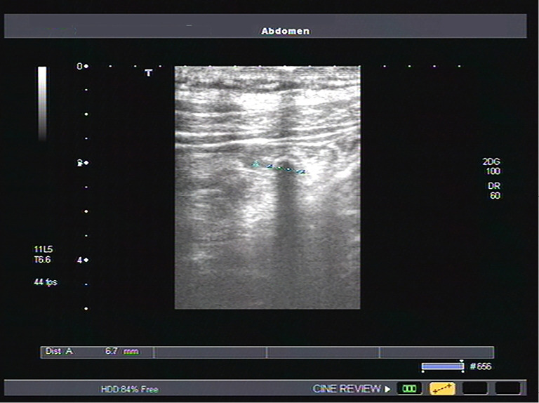
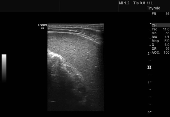
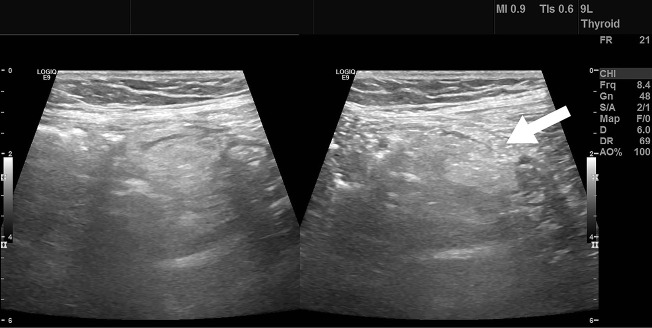
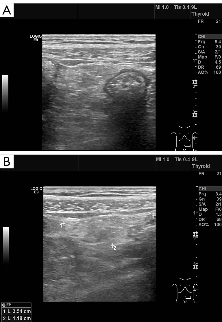
A retrospective analysis was conducted on pediatric patients hospitalized between July 2016 and November 2017 for abdominal pain. Patients were categorized into normal weight, overweight, and obese groups. Conventional and layer-by-layer scanning methods were used by attending physicians to examine abdominal organs, including the liver, gallbladder, spleen, pancreas, kidneys, and bladder. An abdominal probe was employed for rapid screening, followed by a high-frequency probe for detailed three-layer scanning. Ultrasound images were analyzed alongside the children's symptoms and physical signs to provide diagnostic insights.
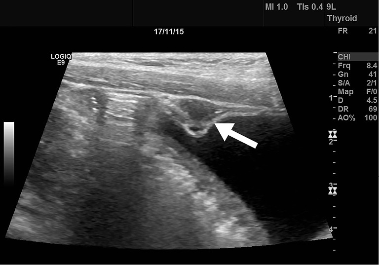
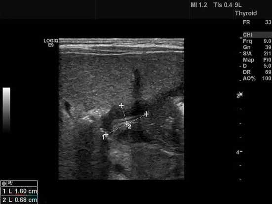
When comparing the conventional and stratified screening groups, several key differences were noted. The stratified group had higher detection rates for mesenteric lymphadenopathy (100% 86%) and peritonitis (94% 27%). Improved detection in the stratified group was due to the identification of peritoneal thickening, leading to higher detection rates for mesenteric fat inflammation (100% 46%), appendicitis (94% 63%), and urachal inflammation (100% 0%). Detection rates for substantial lesions, such as gallstones and ovarian torsion, were similar in both groups (100%). The stratified group also showed significantly better detection of gastrointestinal conditions like gastroenteritis (97% 32%), inguinal hernia (100% 0%), and intestinal ascariasis (100% 47%). Differences in detection rates were observed when abdominal fat layer thickness was between 0.8 and 1.7 cm, with more significant differences when thickness exceeded 1.7 cm.
Real-time ultrasound with stratified screening effectively detects abdominal and pelvic masses, solid organ lesions, and bowel wall thickening, improving disease detection in children, including individuals with increased body mass index. This method is valuable and recommended for wider use.
小儿腹痛是一种常见但诊断具有挑战性的症状,尤其是对于难以表达不适的幼儿。随着肥胖对超声检查准确性的影响日益增加,本研究旨在通过在超声检查中寻找新的途径和方法,特别是在肥胖或超重小儿患者中的应用,来找出小儿腹痛的原因。
对2016年7月至2017年11月因腹痛住院的小儿患者进行回顾性分析。患者分为正常体重、超重和肥胖组。主治医生采用传统和逐层扫描方法检查腹部器官,包括肝脏、胆囊、脾脏、胰腺、肾脏和膀胱。先用腹部探头进行快速筛查,然后用高频探头进行详细的三层扫描。结合儿童的症状和体征对超声图像进行分析,以提供诊断依据。
比较传统筛查组和分层筛查组时,发现了几个关键差异。分层组对肠系膜淋巴结病(100%对86%)和腹膜炎(94%对27%)的检出率更高。分层组检测率提高的原因是发现了腹膜增厚,从而使肠系膜脂肪炎症(100%对46%)、阑尾炎(94%对63%)和脐尿管炎(100%对0%)的检出率更高。两组对胆结石和卵巢扭转等实质性病变的检出率相似(100%)。分层组对肠胃炎(97%对32%)、腹股沟疝(100%对0%)和肠道蛔虫病(100%对47%)等胃肠道疾病的检测也明显更好。当腹部脂肪层厚度在0.8至1.7厘米之间时,检测率存在差异,当厚度超过1.7厘米时差异更显著。
实时超声分层筛查能有效检测腹部和盆腔肿块、实体器官病变及肠壁增厚,提高儿童疾病的检测率,包括体重指数增加的个体。该方法具有重要价值,建议广泛应用。