Yang Yuhao, Tang Mengzhen, Xiong Wangge, Luo Hao, Li Zekun, Yang Xinyu, Chen Weihua, Hu Xiaojing, He Xingdao, Yang Jian
School of Stomatology, Jiangxi Medical College, Nanchang University, Nanchang, China.
Jiangxi Province Key Laboratory of Oral Diseases, Nanchang, China.
Quant Imaging Med Surg. 2025 May 1;15(5):4592-4607. doi: 10.21037/qims-24-2359. Epub 2025 Apr 28.
Oral cancer is the sixth most common cancer worldwide. The detection, prevention, and control of oral potentially malignant disorders (OPMDs) at early stages is imperative to reduce the incidence of oral cancer. This study analyzed ultrastructural and biomechanical tissue properties during tongue cancer development in Sprague-Dawley (SD) rats using optical coherence elastography (OCE). Our investigation examined the changes associated with oral cancer pathogenesis and explored the feasibility of OCE as an early diagnostic tool for oral cancer.
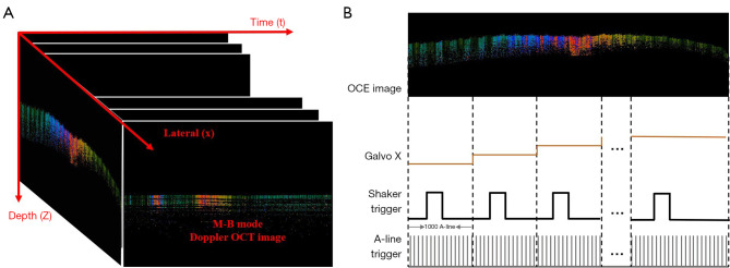
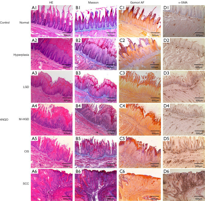
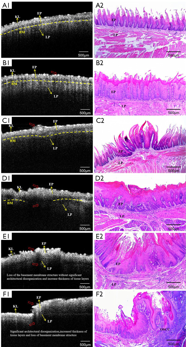
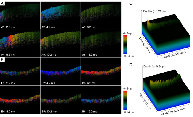
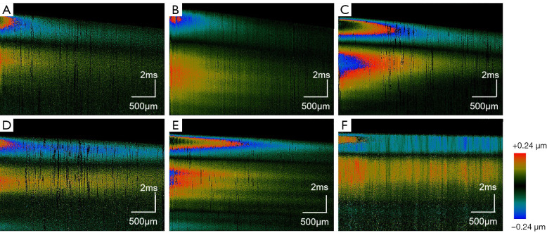
In this study, 4-nitroquinoline-1-oxide (4NQO) was used to induce oral carcinogenesis in SD rats. In total, 10 normal tissues, five hyperplastic lesions, eight low-grade dysplasias (LGDs), eight moderate-high-grade dysplasias (M-HGDs), seven carcinomas in situ (CISs), and seven squamous cell carcinomas (SCCs) were examined. The oral stroma changes were sequentially imaged by shaker-based OCE (shaker-OCE). The changes in the oral stroma from normal to hyperplasia, atypical hyperplasia, CIS, and cancer were determined using OCE, and histological findings such as extracellular matrix (ECM) components (including collagen and elastic fibers) and the expression of cancer-associated fibroblasts (CAFs) were compared at different stages of tongue cancer development.
The findings showed that OCE imaging could be used to accurately distinguish between normal, hyperplasia, atypical hyperplasia, CIS, and oral cancer. Additionally, there were significant differences in the tongue tissue biomechanics across the different lesion levels (P<0.05). Further, as the malignancy of the tongue cancer progressed in the SD rats, the level of collagen fibers gradually increased, showing a positive correlation (r=0.353, P<0.05), while the level of elastic fiber expression gradually decreased, showing a negative correlation (r=-0.776, P<0.05). The alpha-smooth muscle actin (α-SMA) scores of CIS and SCC were statistically significantly higher than those of normal, simple hyperplasia, mild atypical hyperplasia, and moderate-high atypical hyperplasia (P<0.05).
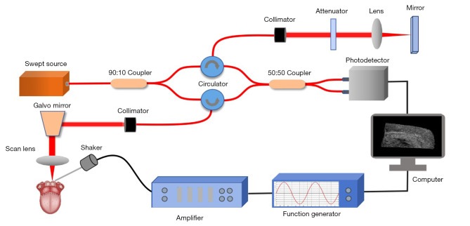
The ability of the shaker-OCE system to obtain the structural and biomechanical characteristics of tongue tissues in a non-invasive, real-time manner was confirmed by this study. It also showed notable benefits in terms of early diagnosis and the dynamic monitoring of tongue cancer. The systematic validation of the physiopathological model revealed a strong correlation between the elastic properties of cancerous tissues and pathological evolution, which provides a theoretical basis and experimental evidence for the clinical application of OCE technology.
口腔癌是全球第六大常见癌症。早期检测、预防和控制口腔潜在恶性疾病(OPMDs)对于降低口腔癌的发病率至关重要。本研究使用光学相干弹性成像(OCE)分析了Sprague-Dawley(SD)大鼠舌癌发生过程中的超微结构和生物力学组织特性。我们的研究考察了与口腔癌发病机制相关的变化,并探讨了OCE作为口腔癌早期诊断工具的可行性。
在本研究中,使用4-硝基喹啉-1-氧化物(4NQO)诱导SD大鼠发生口腔癌。总共检查了10个正常组织、5个增生性病变、8个低级别发育异常(LGDs)、8个中高级别发育异常(M-HGDs)、7个原位癌(CISs)和7个鳞状细胞癌(SCCs)。通过基于振动器的OCE(振动器-OCE)对口腔基质变化进行连续成像。使用OCE确定口腔基质从正常到增生、非典型增生、原位癌和癌症的变化,并比较舌癌发生不同阶段的组织学发现,如细胞外基质(ECM)成分(包括胶原纤维和弹性纤维)以及癌症相关成纤维细胞(CAFs)的表达。
研究结果表明,OCE成像可用于准确区分正常、增生、非典型增生、原位癌和口腔癌。此外,不同病变水平的舌组织生物力学存在显著差异(P<0.05)。此外,随着SD大鼠舌癌恶性程度的进展,胶原纤维水平逐渐升高,呈正相关(r=0.353,P<0.05),而弹性纤维表达水平逐渐降低,呈负相关(r=-0.776,P<0.05)。原位癌和鳞状细胞癌的α-平滑肌肌动蛋白(α-SMA)评分在统计学上显著高于正常、单纯增生、轻度非典型增生和中高度非典型增生(P<0.05)。
本研究证实了振动器-OCE系统以非侵入性、实时方式获取舌组织结构和生物力学特征的能力。它在舌癌的早期诊断和动态监测方面也显示出显著优势。对生理病理模型的系统验证揭示了癌组织弹性特性与病理演变之间的强相关性,为OCE技术的临床应用提供了理论基础和实验证据。