• 文献检索
  • 文档翻译
  • 深度研究
  • 学术资讯
  • Suppr Zotero 插件Zotero 插件
  • 邀请有礼
  • 套餐&价格
  • 历史记录
应用&插件
Suppr Zotero 插件Zotero 插件浏览器插件Mac 客户端Windows 客户端微信小程序
定价
高级版会员购买积分包购买API积分包
服务
文献检索文档翻译深度研究API 文档MCP 服务
关于我们
关于 Suppr公司介绍联系我们用户协议隐私条款
关注我们

Suppr 超能文献

核心技术专利:CN118964589B侵权必究
粤ICP备2023148730 号-1Suppr @ 2026

文献检索

告别复杂PubMed语法,用中文像聊天一样搜索,搜遍4000万医学文献。AI智能推荐,让科研检索更轻松。

立即免费搜索

文件翻译

保留排版,准确专业,支持PDF/Word/PPT等文件格式,支持 12+语言互译。

免费翻译文档

深度研究

AI帮你快速写综述,25分钟生成高质量综述,智能提取关键信息,辅助科研写作。

立即免费体验

超声弹性成像

Ultrasound elastography.

作者信息

Cui Xin-Wu, Li Kang-Ning, Yi Ai-Jiao, Wang Bin, Wei Qi, Wu Ge-Ge, Dietrich Christoph F

机构信息

Department of Medical Ultrasound, Tongji Hospital, Tongji Medical College, Huazhong University of Science and Technology, Wuhan, Hubei Province, China.

Department of Ultrasound, The First People's Hospital of Yueyang, Yueyang, Hunan Province, China.

出版信息

Endosc Ultrasound. 2022 Jul-Aug;11(4):252-274. doi: 10.4103/EUS-D-21-00151.

DOI:10.4103/EUS-D-21-00151
PMID:35532576
原文链接:https://pmc.ncbi.nlm.nih.gov/articles/PMC9526103/
Abstract

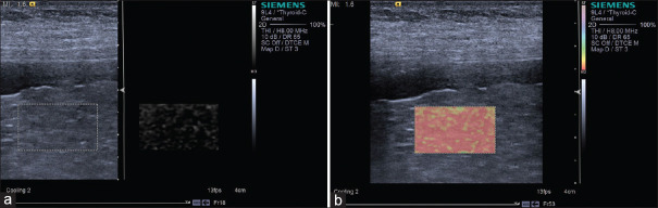
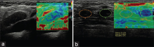
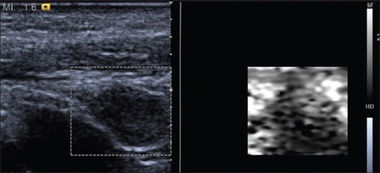
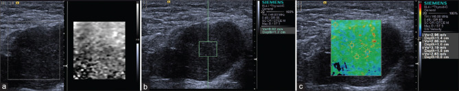
Physicians have used palpation as a diagnostic examination to understand the elastic properties of pathology for a long time since they realized that tissue stiffness is closely related to its biological characteristics. US elastography provided new diagnostic information about elasticity comparing with the morphological feathers of traditional US, and thus expanded the scope of the application in clinic. US elastography is now widely used in the field of diagnosis and differential diagnosis of abnormality, evaluating the degree of fibrosis and assessment of treatment response for a range of diseases. The World Federation of Ultrasound Medicine and Biology divided elastographic techniques into strain elastography (SE), transient elastography and acoustic radiation force impulse (ARFI). The ARFI techniques can be further classified into point shear wave elastography (SWE), 2D SWE, and 3D SWE techniques. The SE measures the strain, while the shear wave-based techniques (including TE and ARFI techniques) measure the speed of shear waves in tissues. In this review, we discuss the various techniques separately based on their basic principles, clinical applications in various organs, and advantages and limitations and which might be most appropriate given that the majority of doctors have access to only one kind of machine.

摘要

自从医生们认识到组织硬度与其生物学特性密切相关以来,他们长期以来一直将触诊作为一种诊断检查方法来了解病变的弹性特性。与传统超声的形态学特征相比,超声弹性成像提供了有关弹性的新诊断信息,从而扩大了其临床应用范围。目前,超声弹性成像广泛应用于各种疾病的异常诊断与鉴别诊断、纤维化程度评估以及治疗反应评估等领域。世界超声医学与生物学联合会将弹性成像技术分为应变弹性成像(SE)、瞬时弹性成像和声学辐射力脉冲(ARFI)。ARFI技术可进一步分为点剪切波弹性成像(SWE)、二维SWE和三维SWE技术。SE测量应变,而基于剪切波的技术(包括TE和ARFI技术)测量组织中的剪切波速度。在本综述中,我们将根据各种技术的基本原理、在各个器官中的临床应用、优缺点,以及鉴于大多数医生只能使用一种机器的情况下哪种技术可能最合适,分别对这些技术进行讨论。

https://cdn.ncbi.nlm.nih.gov/pmc/blobs/5b91/9526103/c0aa066ba1d2/EUS-11-252-g029.jpg
https://cdn.ncbi.nlm.nih.gov/pmc/blobs/5b91/9526103/60b859e31ed4/EUS-11-252-g001.jpg
https://cdn.ncbi.nlm.nih.gov/pmc/blobs/5b91/9526103/34a49ff52017/EUS-11-252-g007.jpg
https://cdn.ncbi.nlm.nih.gov/pmc/blobs/5b91/9526103/4c53991a2c4e/EUS-11-252-g010.jpg
https://cdn.ncbi.nlm.nih.gov/pmc/blobs/5b91/9526103/9d53376a51f0/EUS-11-252-g011.jpg
https://cdn.ncbi.nlm.nih.gov/pmc/blobs/5b91/9526103/5548cbc85263/EUS-11-252-g012.jpg
https://cdn.ncbi.nlm.nih.gov/pmc/blobs/5b91/9526103/1ed98d9acee9/EUS-11-252-g013.jpg
https://cdn.ncbi.nlm.nih.gov/pmc/blobs/5b91/9526103/873c471b2994/EUS-11-252-g014.jpg
https://cdn.ncbi.nlm.nih.gov/pmc/blobs/5b91/9526103/08c832c36f72/EUS-11-252-g015.jpg
https://cdn.ncbi.nlm.nih.gov/pmc/blobs/5b91/9526103/715a8823876b/EUS-11-252-g016.jpg
https://cdn.ncbi.nlm.nih.gov/pmc/blobs/5b91/9526103/a8c5f5f12232/EUS-11-252-g017.jpg
https://cdn.ncbi.nlm.nih.gov/pmc/blobs/5b91/9526103/d6c2b8f0b9e4/EUS-11-252-g018.jpg
https://cdn.ncbi.nlm.nih.gov/pmc/blobs/5b91/9526103/8182e34f7c0e/EUS-11-252-g019.jpg
https://cdn.ncbi.nlm.nih.gov/pmc/blobs/5b91/9526103/b69e220a0da3/EUS-11-252-g020.jpg
https://cdn.ncbi.nlm.nih.gov/pmc/blobs/5b91/9526103/c891f90eea77/EUS-11-252-g021.jpg
https://cdn.ncbi.nlm.nih.gov/pmc/blobs/5b91/9526103/55b86dad700b/EUS-11-252-g022.jpg
https://cdn.ncbi.nlm.nih.gov/pmc/blobs/5b91/9526103/f002bab93a19/EUS-11-252-g023.jpg
https://cdn.ncbi.nlm.nih.gov/pmc/blobs/5b91/9526103/8744e44ab5e4/EUS-11-252-g024.jpg
https://cdn.ncbi.nlm.nih.gov/pmc/blobs/5b91/9526103/d1e9a987937a/EUS-11-252-g025.jpg
https://cdn.ncbi.nlm.nih.gov/pmc/blobs/5b91/9526103/c646863a1d83/EUS-11-252-g026.jpg
https://cdn.ncbi.nlm.nih.gov/pmc/blobs/5b91/9526103/9b08963ce4d3/EUS-11-252-g027.jpg
https://cdn.ncbi.nlm.nih.gov/pmc/blobs/5b91/9526103/c2143000f635/EUS-11-252-g028.jpg
https://cdn.ncbi.nlm.nih.gov/pmc/blobs/5b91/9526103/c0aa066ba1d2/EUS-11-252-g029.jpg

相似文献

1
Ultrasound elastography.超声弹性成像
Endosc Ultrasound. 2022 Jul-Aug;11(4):252-274. doi: 10.4103/EUS-D-21-00151.
2
Ultrasound Shear Wave Elastography for Liver Disease. A Critical Appraisal of the Many Actors on the Stage.用于肝脏疾病的超声剪切波弹性成像:对该领域众多参与者的批判性评估
Ultraschall Med. 2016 Feb;37(1):1-5. doi: 10.1055/s-0035-1567037. Epub 2016 Feb 12.
3
Comparison of strain elastography, point shear wave elastography using acoustic radiation force impulse imaging and 2D-shear wave elastography for the differentiation of thyroid nodules.应变弹性成像、声辐射力脉冲成像点剪切波弹性成像与二维剪切波弹性成像在甲状腺结节鉴别诊断中的比较。
PLoS One. 2018 Sep 17;13(9):e0204095. doi: 10.1371/journal.pone.0204095. eCollection 2018.
4
Assessment of liver fibrosis with 2-D shear wave elastography in comparison to transient elastography and acoustic radiation force impulse imaging in patients with chronic liver disease.二维剪切波弹性成像评估慢性肝病患者肝纤维化,并与瞬时弹性成像和声辐射力脉冲成像进行比较。
Ultrasound Med Biol. 2015 Sep;41(9):2350-9. doi: 10.1016/j.ultrasmedbio.2015.04.014. Epub 2015 Jun 24.
5
Acoustic radiation force impulse induced strain elastography and point shear wave elastography for evaluation of thyroid nodules.声辐射力脉冲诱导应变弹性成像和点剪切波弹性成像在甲状腺结节评估中的应用
Int J Clin Exp Med. 2015 Jul 15;8(7):10956-63. eCollection 2015.
6
Value of Real-Time Shear Wave Elastography Versus Acoustic Radiation Force Impulse Imaging in the Diagnosis of Female Bladder Neck Obstruction.实时剪切波弹性成像与声辐射力脉冲成像在女性膀胱颈梗阻诊断中的价值。
J Ultrasound Med. 2019 Sep;38(9):2427-2435. doi: 10.1002/jum.14941. Epub 2019 Jan 25.
7
The diagnosis value of acoustic radiation force impulse (ARFI) elastography for thyroid malignancy without highly suspicious features on conventional ultrasound.声辐射力脉冲(ARFI)弹性成像对常规超声无高度可疑特征的甲状腺恶性肿瘤的诊断价值。
Int J Clin Exp Med. 2015 Sep 15;8(9):15362-72. eCollection 2015.
8
Initial Experience With Ultrasound Elastography for Diagnosis of Major Salivary Gland Lesions.超声弹性成像诊断大唾液腺病变的初步经验
J Ultrasound Med. 2016 Dec;35(12):2597-2606. doi: 10.7863/ultra.15.11093. Epub 2016 Oct 25.
9
Comparison of three ultrasound based elastographic techniques in children and adolescents with chronic diffuse liver diseases.三种基于超声的弹性成像技术在儿童和青少年慢性弥漫性肝病中的比较。
Med Ultrason. 2016 Jun;18(2):145-50. doi: 10.11152/mu.2013.2066.182.bet.
10
Shear wave elastography of the testes in a healthy study collective - Differences in standard values between ARFI and VTIQ techniques.健康研究群体中睾丸的剪切波弹性成像——ARFI和VTIQ技术标准值的差异
Clin Hemorheol Microcirc. 2016;64(4):721-728. doi: 10.3233/CH-168052.

引用本文的文献

1
B-Mode Ultrasound May Be an Early Marker in Acute Kidney Injury.B超可能是急性肾损伤的早期标志物。
Diagnostics (Basel). 2025 Aug 14;15(16):2034. doi: 10.3390/diagnostics15162034.
2
Plaque biomarkers from high-frame rate vector flow imaging and shear wave elastography in mild carotid stenosis.高帧率矢量血流成像和剪切波弹性成像在轻度颈动脉狭窄中的斑块生物标志物
Ultrasonography. 2025 Jul;44(4):274-284. doi: 10.14366/usg.25031. Epub 2025 May 6.
3
From imaging techniques to laboratory tests: a multimodal stratification for varicocele-associated decreased semen quality.
从成像技术到实验室检测:精索静脉曲张相关性精液质量下降的多模态分层
Basic Clin Androl. 2025 Jun 2;35(1):20. doi: 10.1186/s12610-025-00268-z.
4
Can transabdominal shear wave elastography play a role in solving the dilemma of complex cystic and solid ovarian tumors by ultrasound?经腹剪切波弹性成像在解决超声诊断复杂囊性及实性卵巢肿瘤的困境中能发挥作用吗?
J Ultrasound. 2025 May 31. doi: 10.1007/s40477-025-01027-6.
5
Sleep behaviors and metabolic-associated fatty liver disease.睡眠行为与代谢相关脂肪性肝病
PLoS One. 2025 May 28;20(5):e0323715. doi: 10.1371/journal.pone.0323715. eCollection 2025.
6
Utilizing Shear Wave Elastography for the Evaluation of Ocular Involvement in Systemic Sclerosis.利用剪切波弹性成像技术评估系统性硬化症的眼部受累情况。
Diagnostics (Basel). 2025 May 13;15(10):1227. doi: 10.3390/diagnostics15101227.
7
Quantitative assessment of biomechanical changes in oral lesions at different cancerous stages using optical coherence elastography.使用光学相干弹性成像技术对口腔病变在不同癌变阶段的生物力学变化进行定量评估。
Quant Imaging Med Surg. 2025 May 1;15(5):4592-4607. doi: 10.21037/qims-24-2359. Epub 2025 Apr 28.
8
B-Mode Ultrasonography and Acoustic Radiation Force Impulse Elastography in Evaluation of Urothelial Carcinoma in Dogs.B 型超声检查和声学辐射力脉冲弹性成像在犬膀胱移行细胞癌评估中的应用
Animals (Basel). 2025 Apr 26;15(9):1223. doi: 10.3390/ani15091223.
9
Elastography Enhances the Diagnostic Performance of Conventional Ultrasonography in Differentiating Benign from Malignant Superficial Lymphadenopathies.弹性成像提高了传统超声在鉴别浅表淋巴结病变良恶性方面的诊断性能。
Cancers (Basel). 2025 Apr 28;17(9):1480. doi: 10.3390/cancers17091480.
10
Advances in Skin Ultrasonography for Malignant and Benign Tumors of the Head and Neck: Current Insights and Future Directions.头颈部恶性和良性肿瘤的皮肤超声检查进展:当前见解与未来方向
J Clin Med. 2025 Mar 27;14(7):2298. doi: 10.3390/jcm14072298.