Ahmadzadeh Chaleshtori Mohammad Amin, Salehzadeh Ali, Peymani Maryam
Department of Biology, Rasht Branch, Islamic Azad University, Rasht, Iran.
Department of Biology, ShK. C., Islamic Azad University, Shahrekord, Iran.
Sci Rep. 2025 May 20;15(1):17467. doi: 10.1038/s41598-025-02189-3.
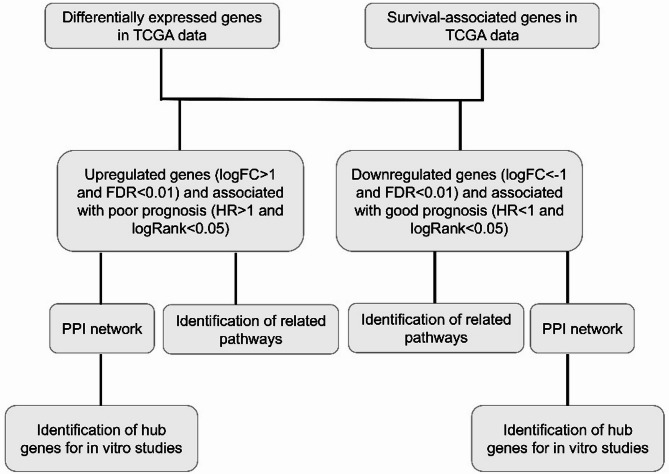
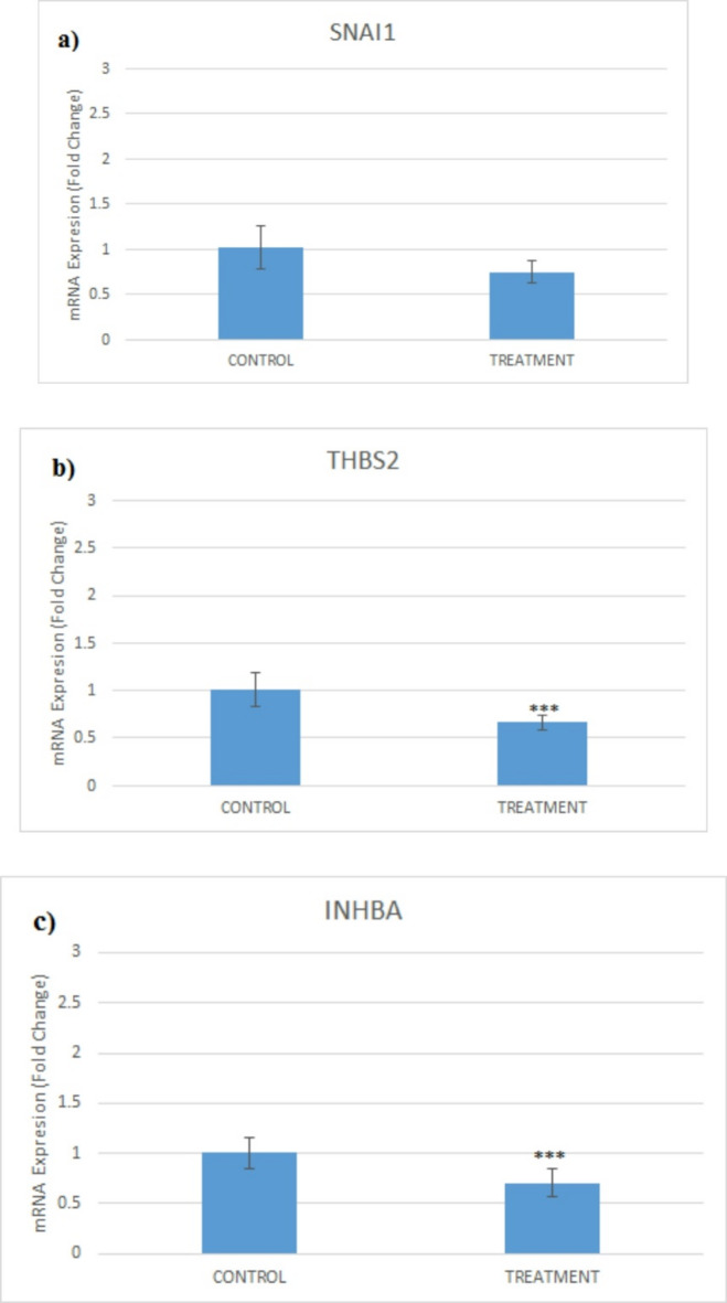
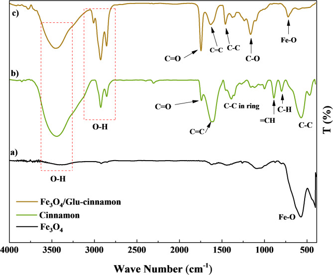
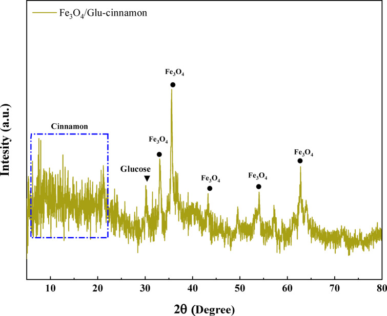
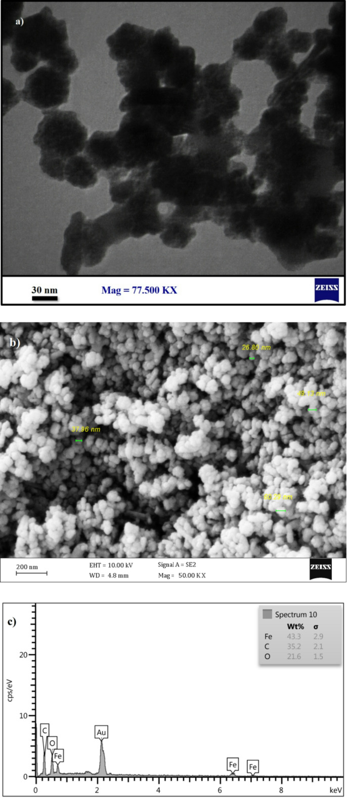
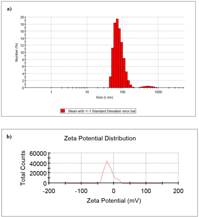
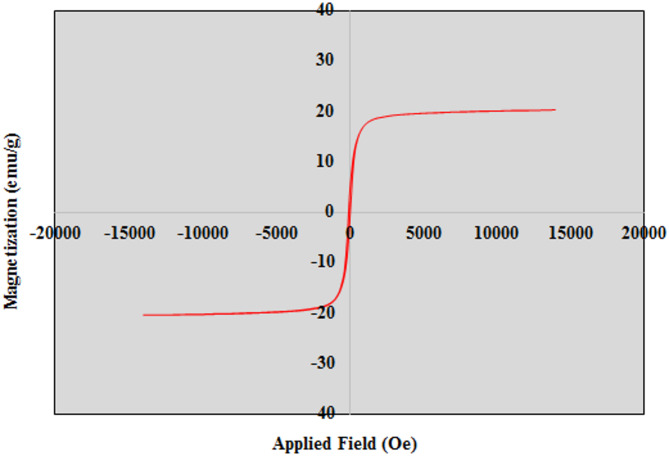
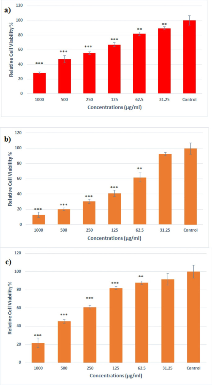
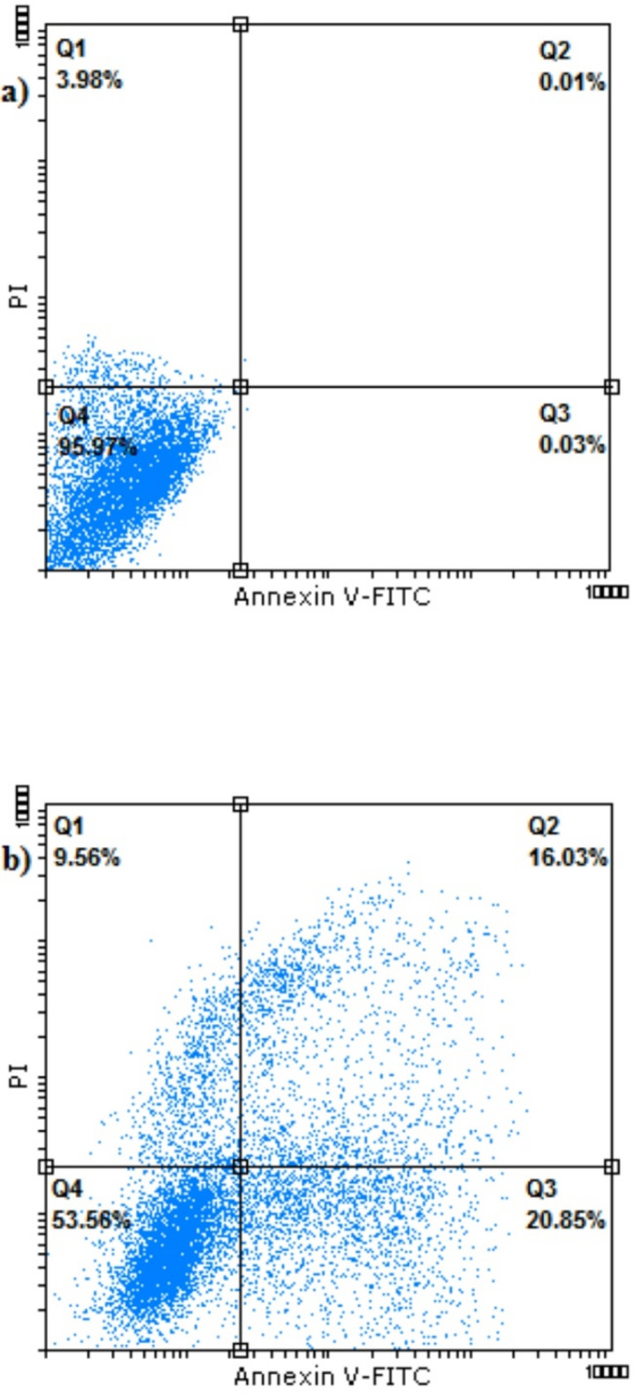
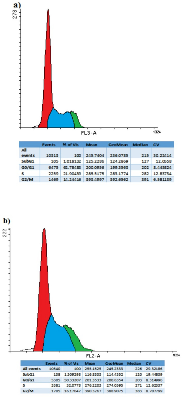
Finding the molecular targets involved in the severity and drug resistance of Colorectal cancer (CRC) and applying targeted treatments against them is a promising approach. In this study, some candidate oncogenes related to disease severity and mortality were identified by extracting bioinformatics data, and the effect of FeO@Glu-Cinnamon NPs on the survival of CRC cells (SW480) and the expression of these oncogenes was investigated. The NPs were characterized by FT-IR, XRD, DLS and zeta potential measurement, TEM and SEM imaging, EDS-mapping and VSM analysis. Cytotoxicity of the NPs was evaluated by the MTT assay and a flow cytometry analysis was done to investigate cell apoptosis/necrosis levels and cell cycle analysis of cancer cells. The FeO@Glu-Cinnamon NPs with spherical morphology were correctly synthesized, containing no elemental impurities, with a size range of 26.8-60.2 nm, DLS of 213 nm, zeta potential of -15.4mV and maximum magnetization level of 20.33emu/g. Treatment of cancer cells with the NPs elevated primary and late apoptosis and cell necrosis levels to 20.85, 16.83 and 9.56% and treated cells were mainly arrested at the S and G2/M phases. The expression level of the oncogenes associated with mortality, SNAI1, THBS2 and INHBA reduced to 0.74, 0.66 and 0.7 folds, respectively. The magnetic properties of FeO NPs enable their potential use in targeted drug delivery, allowing for site-specific accumulation within tumors. This could minimize systemic toxicity while enhancing treatment efficacy.
寻找参与结直肠癌(CRC)严重程度和耐药性的分子靶点并针对这些靶点进行靶向治疗是一种很有前景的方法。在本研究中,通过提取生物信息学数据确定了一些与疾病严重程度和死亡率相关的候选癌基因,并研究了FeO@Glu-肉桂纳米颗粒对CRC细胞(SW480)存活以及这些癌基因表达的影响。通过傅里叶变换红外光谱(FT-IR)、X射线衍射(XRD)、动态光散射(DLS)和zeta电位测量、透射电子显微镜(TEM)和扫描电子显微镜(SEM)成像、能谱映射(EDS-mapping)和振动样品磁强计(VSM)分析对纳米颗粒进行了表征。通过MTT法评估纳米颗粒的细胞毒性,并进行流式细胞术分析以研究癌细胞的凋亡/坏死水平和细胞周期分析。成功合成了具有球形形态的FeO@Glu-肉桂纳米颗粒,不含元素杂质,尺寸范围为26.8-60.2nm,DLS为213nm,zeta电位为-15.4mV,最大磁化强度为20.33emu/g。用纳米颗粒处理癌细胞后,早期和晚期凋亡以及细胞坏死水平分别提高到20.85%、16.83%和9.56%,处理后的细胞主要停滞在S期和G2/M期。与死亡率相关的癌基因SNAI1、THBS2和INHBA的表达水平分别降至0.74、0.66和0.7倍。FeO纳米颗粒的磁性使其有可能用于靶向药物递送,从而在肿瘤内实现位点特异性积累。这可以在提高治疗效果的同时将全身毒性降至最低。