• 文献检索
  • 文档翻译
  • 深度研究
  • 学术资讯
  • Suppr Zotero 插件Zotero 插件
  • 邀请有礼
  • 套餐&价格
  • 历史记录
应用&插件
Suppr Zotero 插件Zotero 插件浏览器插件Mac 客户端Windows 客户端微信小程序
定价
高级版会员购买积分包购买API积分包
服务
文献检索文档翻译深度研究API 文档MCP 服务
关于我们
关于 Suppr公司介绍联系我们用户协议隐私条款
关注我们

Suppr 超能文献

核心技术专利:CN118964589B侵权必究
粤ICP备2023148730 号-1Suppr @ 2026

文献检索

告别复杂PubMed语法,用中文像聊天一样搜索,搜遍4000万医学文献。AI智能推荐,让科研检索更轻松。

立即免费搜索

文件翻译

保留排版,准确专业,支持PDF/Word/PPT等文件格式,支持 12+语言互译。

免费翻译文档

深度研究

AI帮你快速写综述,25分钟生成高质量综述,智能提取关键信息,辅助科研写作。

立即免费体验

氩激光光凝术后糖尿病视网膜的视网膜功能:视网膜电图评估

Retinal function of the diabetic retina after argon laser photocoagulation assessed electroretinographically.

作者信息

Perlman I, Gdal-On M, Miller B, Zonis S

出版信息

Br J Ophthalmol. 1985 Apr;69(4):240-6. doi: 10.1136/bjo.69.4.240.

DOI:10.1136/bjo.69.4.240
PMID:4039601
原文链接:https://pmc.ncbi.nlm.nih.gov/articles/PMC1040575/
Abstract

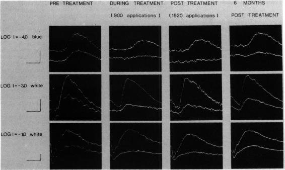
Electroretinographic (ERG) responses were measured in diabetic patients before, during, and after panretinal photocoagulation treatment with argon laser. The laser applications reduced considerably the amplitudes of the a and b waves of the ERG. Moreover, the relationship between the amplitude of the b wave and that of the a wave was severely affected, resulting in ERG responses of abnormal pattern. The b waves were smaller than expected from the a waves. These findings indicated that the photocoagulation treatment not only destroyed the retinal areas directly illuminated by the laser beam, but also affected the functional integrity of adjacent areas. These additional effects resulted in subnormal signal transmission from the photoreceptors to the proximal retina.

摘要

对糖尿病患者在氩激光全视网膜光凝治疗前、治疗期间和治疗后进行了视网膜电图(ERG)反应测量。激光照射使ERG的a波和b波振幅大幅降低。此外,b波振幅与a波振幅之间的关系受到严重影响,导致ERG反应模式异常。b波比根据a波预期的要小。这些发现表明,光凝治疗不仅破坏了激光束直接照射的视网膜区域,还影响了相邻区域的功能完整性。这些额外的影响导致从光感受器到视网膜近端的信号传递低于正常水平。

https://cdn.ncbi.nlm.nih.gov/pmc/blobs/77c7/1040575/13a9529b5619/brjopthal00136-0006-a.jpg

相似文献

1
Retinal function of the diabetic retina after argon laser photocoagulation assessed electroretinographically.氩激光光凝术后糖尿病视网膜的视网膜功能:视网膜电图评估
Br J Ophthalmol. 1985 Apr;69(4):240-6. doi: 10.1136/bjo.69.4.240.
2
Electrophysiological studies before and after argon-laser photocoagulation in diabetic retinopathy.糖尿病视网膜病变患者氩激光光凝治疗前后的电生理研究
Ophthalmologica. 1977;176(3):133-44. doi: 10.1159/000308705.
3
Electroretinographic findings in panretinal photocoagulation for diabetic retinopathy. A randomized study with blue-green argon and red krypton lasers.糖尿病性视网膜病变全视网膜光凝的视网膜电图研究。一项关于蓝绿氩激光和红氪激光的随机研究。
Graefes Arch Clin Exp Ophthalmol. 1990;228(3):232-6. doi: 10.1007/BF00920026.
4
Comparative electroretinograms in argon laser and xenon arc panretinal photocoagulation.氩激光与氙弧全视网膜光凝的比较视网膜电图
Br J Ophthalmol. 1983 Aug;67(8):520-5. doi: 10.1136/bjo.67.8.520.
5
Recovery of the electroretinogram in rabbits after argon laser photocoagulation.氩激光光凝后兔视网膜电图的恢复情况
Invest Ophthalmol Vis Sci. 1987 Sep;28(9):1605-13.
6
An electrophysiological study on panretinal photocoagulation in diabetic retinopathy.糖尿病视网膜病变全视网膜光凝的电生理研究
Metab Pediatr Syst Ophthalmol (1985). 1987;10(3):64-7.
7
[The effects of photocoagulation on the electrical activity of the diabetic retina].
Oftalmologia. 2000;51(2):29-34.
8
Visual fields and electroretinography following extensive photocoagulation.广泛光凝后的视野和视网膜电图
Arch Ophthalmol. 1975 Aug;93(8):591-8. doi: 10.1001/archopht.1975.01010020575004.
9
The electroretinogram after peripheral retinal ablation in diabetic retinopathy.糖尿病视网膜病变周边视网膜消融后的视网膜电图。
Am J Ophthalmol. 1976 Apr;81(4):397-402. doi: 10.1016/0002-9394(76)90293-2.
10
ERG and VER findings after laser photocoagulation of the retina.视网膜激光光凝术后的视网膜电图(ERG)和视觉诱发电位(VER)检查结果。
Metab Pediatr Syst Ophthalmol. 1982;6(2):101-5.

引用本文的文献

1
Comparison of functional changes of retina after subthreshold and threshold pan-retinal photocoagulation in severe non-proliferative diabetic retinopathy.严重非增生型糖尿病视网膜病变患者经阈下和阈值全视网膜光凝后视网膜功能变化的比较。
Lasers Med Sci. 2022 Dec;37(9):3561-3569. doi: 10.1007/s10103-022-03635-8. Epub 2022 Sep 7.
2
Clinical electroretinography in diabetic retinopathy: a review.临床视网膜电图在糖尿病视网膜病变中的应用:综述。
Surv Ophthalmol. 2022 May-Jun;67(3):712-722. doi: 10.1016/j.survophthal.2021.08.011. Epub 2021 Sep 4.
3
Electroretinogram Changes Following Sequential Panretinal Photocoagulation for Proliferative Diabetic Retinopathy.

本文引用的文献

1
Comparative electroretinograms in argon laser and xenon arc panretinal photocoagulation.氩激光与氙弧全视网膜光凝的比较视网膜电图
Br J Ophthalmol. 1983 Aug;67(8):520-5. doi: 10.1136/bjo.67.8.520.
2
Relationship between the amplitudes of the b wave and the a wave as a useful index for evaluating the electroretinogram.
Br J Ophthalmol. 1983 Jul;67(7):443-8. doi: 10.1136/bjo.67.7.443.
3
Photocoagulation in diabetic retinopathy.
Am J Ophthalmol. 1971 Aug;72(2):299-321. doi: 10.1016/0002-9394(71)91300-6.
4
增殖性糖尿病视网膜病变行序贯性全视网膜光凝术后的视网膜电图变化
Clin Ophthalmol. 2020 Mar 30;14:967-975. doi: 10.2147/OPTH.S248678. eCollection 2020.
4
Electroretinogram evaluation for the treatment of proliferative diabetic retinopathy by short-pulse pattern scanning laser panretinal photocoagulation.短脉冲模式扫描激光全视网膜光凝治疗增殖性糖尿病视网膜病变的视网膜电图评估
Lasers Med Sci. 2018 Jul;33(5):1095-1102. doi: 10.1007/s10103-018-2474-0. Epub 2018 Mar 15.
5
Photopic full-field electroretinography and optical coherence tomography in type 1 diabetic retinopathy.1型糖尿病视网膜病变的明视全视野视网膜电图和光学相干断层扫描
Graefes Arch Clin Exp Ophthalmol. 2015 Jul;253(7):989-97. doi: 10.1007/s00417-015-3034-y. Epub 2015 May 26.
6
Electroretinographic findings associated with panretinal photocoagulation (PRP) versus PRP plus intravitreal ranibizumab treatment for high-risk proliferative diabetic retinopathy.全视网膜光凝(PRP)与PRP联合玻璃体内注射雷珠单抗治疗高危增殖性糖尿病视网膜病变的视网膜电图检查结果
Doc Ophthalmol. 2012 Jun;124(3):225-36. doi: 10.1007/s10633-012-9322-5. Epub 2012 Mar 29.
7
Alteration of melatonin secretion in patients with type 2 diabetes and proliferative diabetic retinopathy.2型糖尿病合并增殖性糖尿病视网膜病变患者褪黑素分泌的改变。
Clin Ophthalmol. 2011;5:655-60. doi: 10.2147/OPTH.S19559. Epub 2011 May 19.
8
Macular function assessed with mfERG before and after panretinal photocoagulation in patients with proliferative diabetic retinopathy.增殖性糖尿病视网膜病变患者全视网膜光凝治疗前后,采用多焦视网膜电图评估黄斑功能。
Doc Ophthalmol. 2004 Sep;109(2):115-21. doi: 10.1007/s10633-004-4862-y.
9
Visual function in hypermetropia. An electroretinographic and psychophysical study.
Doc Ophthalmol. 1993;84(1):47-59. doi: 10.1007/BF01203282.
10
Pattern electroretinograms become abnormal when background diabetic retinopathy deteriorates to a preproliferative stage: possible use as a screening test.当背景性糖尿病视网膜病变恶化至增殖前期时,图形视网膜电图会变得异常:可能用作一种筛查测试。
Br J Ophthalmol. 1986 May;70(5):330-5. doi: 10.1136/bjo.70.5.330.
Visual fields and electroretinography following extensive photocoagulation.广泛光凝后的视野和视网膜电图
Arch Ophthalmol. 1975 Aug;93(8):591-8. doi: 10.1001/archopht.1975.01010020575004.
5
The electroretinogram after peripheral retinal ablation in diabetic retinopathy.糖尿病视网膜病变周边视网膜消融后的视网膜电图。
Am J Ophthalmol. 1976 Apr;81(4):397-402. doi: 10.1016/0002-9394(76)90293-2.
6
The influence of photocoagulation of the retina on the electroretinogram.视网膜光凝对视网膜电图的影响。
Doc Ophthalmol. 1977 Feb 28;42(2):369-73. doi: 10.1007/BF02742250.
7
Electrophysiological studies before and after argon-laser photocoagulation in diabetic retinopathy.糖尿病视网膜病变患者氩激光光凝治疗前后的电生理研究
Ophthalmologica. 1977;176(3):133-44. doi: 10.1159/000308705.