Stricker Priscila Elias Ferreira, de Oliveira Nathalia Barth, Mogharbel Bassam Felipe, Irioda Ana Carolina, da Rosa Nádia Nascimento, Lührs Larissa, Saçaki Claudia Sayuri, Munhoz da Rocha Isadora, Alves Lysangela Ronalte, Poubel Saloe Bispo, Cardoso da Silva Julia, Carvalho Paulo Costa, Fischer Juliana Saldanha da Gama, de Carvalho Katherine Athayde Teixeira
Pelé Pequeno Príncipe Research Institute, Child and Adolescent Health Research & Pequeno Príncipe Faculties, Advanced Therapy and Cellular Biotechnology in Regenerative Medicine Department, Curitiba, PR, Brazil.
Gene Expression Regulation Laboratory, Carlos Chagas Institute, FIOCRUZ, Curitiba, PR, Brazil.
Int J Nanomedicine. 2025 May 25;20:6675-6699. doi: 10.2147/IJN.S502031. eCollection 2025.
The therapeutic effect of stem cells is attributed to their direct maturation into somatic cells and their paracrine effects, which influence the extracellular environment. One such component released is extracellular vesicles containing proteins and genetic materials with immunomodulatory functions and facilitating cell-to-cell communication.
The study's main objective was to characterize extracellular vesicles (EVs) from Human Neural Precursor Cells (hNPCs).
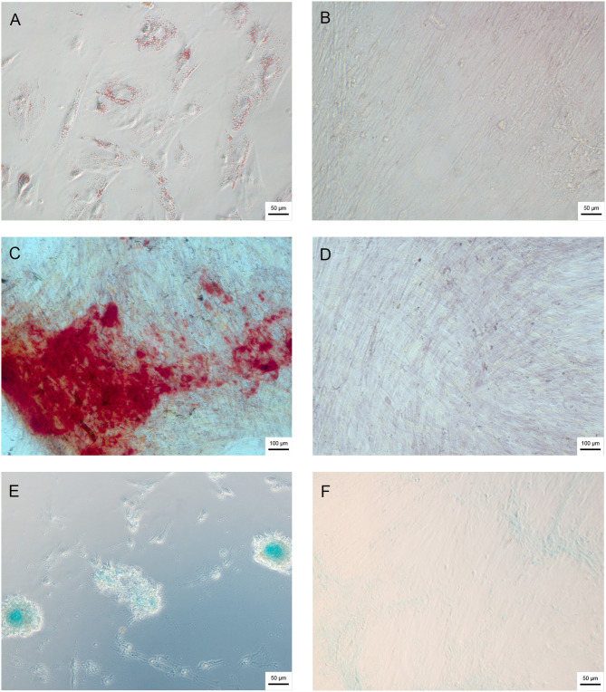
Wharton's Jelly mesenchymal stem cells (WJ-MSCs) were isolated by explant technique and characterized by flow cytometry and trilineage differentiation. The hNPCs obtained from neurospheres were produced by seeding WJ-MSCs on a natural functional biopolymer matrix. EVs derived from WJ-MSCs and hNPCs were isolated by precipitation methodology and characterized by flow cytometry, nanoparticle tracking analysis (NTA), scanning electron microscopy (TEM), and proteomic.
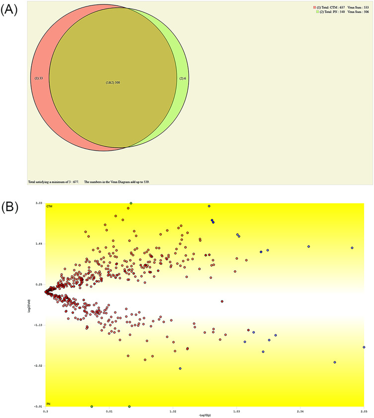
hNPCs expressed proteins and genes characteristic of neural precursor cells. The EVs were characterized by flow cytometry and showed varied expression for the markers CD63, CD9, and CD81, indicating different subpopulations based on their origin of formation. NTA and TEM of the EVs exhibited characteristic size, shape, and structural integrity consistent with the criteria established by the International Society for Extracellular Vesicles (ISEV). EV-hNPCs function enrichment analysis of the proteomic results showed that these vesicles presented abundant proteins directly involved in neuronal biological processes such as plasticity, transduction, postsynaptic density, and overall brain development.
The results indicate that EVs derived from hNPCs maintain key neural precursor characteristics and exhibit marker variability, suggesting distinct subpopulations. Their structural integrity aligns with ISEV standards, supporting their potential as reliable biological entities. The proteomic analysis highlights their role in neuronal functions, reinforcing their applicability in neurodegenerative research and therapeutic strategies.
The EVs were successfully isolated from hNPCs with abundant proteins involved in neuronal processes, making them attractive for acellular therapies to treat neurodegenerative diseases.
干细胞的治疗效果归因于其直接成熟为体细胞及其旁分泌作用,这些旁分泌作用会影响细胞外环境。释放的一种此类成分是细胞外囊泡,其含有具有免疫调节功能并促进细胞间通讯的蛋白质和遗传物质。
本研究的主要目的是对来自人神经前体细胞(hNPCs)的细胞外囊泡(EVs)进行表征。
采用组织块培养技术分离脐带华通氏胶间充质干细胞(WJ-MSCs),并通过流式细胞术和三系分化进行表征。通过将WJ-MSCs接种在天然功能性生物聚合物基质上产生从神经球获得的hNPCs。通过沉淀法分离源自WJ-MSCs和hNPCs的EVs,并通过流式细胞术、纳米颗粒跟踪分析(NTA)、扫描电子显微镜(TEM)和蛋白质组学进行表征。
hNPCs表达神经前体细胞的特征性蛋白质和基因。通过流式细胞术对EVs进行表征,结果显示其标志物CD63、CD9和CD81表达各异,表明基于其形成来源存在不同亚群。EVs的NTA和TEM显示出与国际细胞外囊泡协会(ISEV)制定的标准一致的特征尺寸、形状和结构完整性。蛋白质组学结果的EV-hNPCs功能富集分析表明,这些囊泡呈现出大量直接参与神经元生物学过程的蛋白质,如可塑性、转导、突触后密度和整体脑发育。
结果表明,源自hNPCs的EVs保持关键的神经前体特征并表现出标志物变异性,提示存在不同亚群。其结构完整性符合ISEV标准,支持其作为可靠生物实体的潜力。蛋白质组学分析突出了它们在神经元功能中的作用,加强了它们在神经退行性疾病研究和治疗策略中的适用性。
成功从hNPCs中分离出EVs,其含有大量参与神经元过程的蛋白质,使其在治疗神经退行性疾病的无细胞疗法中具有吸引力。