Sakano Yuri, Kawasaki Hideya
NanoSuit Research Laboratory, Division of Preeminent Bioimaging Research, Institute of Photonics Medicine, Hamamatsu University School of Medicine, Hamamatsu, Shizuoka, Japan.
Immun Inflamm Dis. 2025 Jun;13(6):e70212. doi: 10.1002/iid3.70212.
Molluscum contagiosum (MC) is a common viral skin infection caused by members of the Poxviridae family. It primarily affects children, sexually active adults, and immunocompromised individuals. Although MC spreads through direct contact and auto-inoculation, the precise mechanisms by which the virus penetrates the skin barrier remain poorly understood.
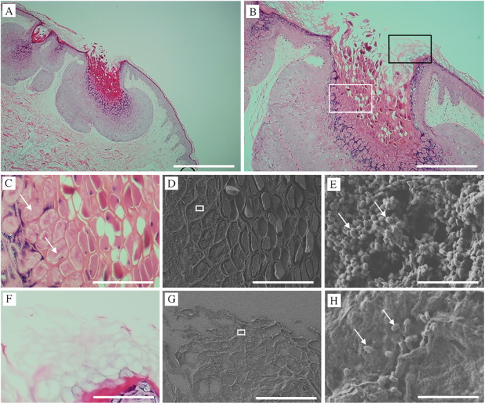
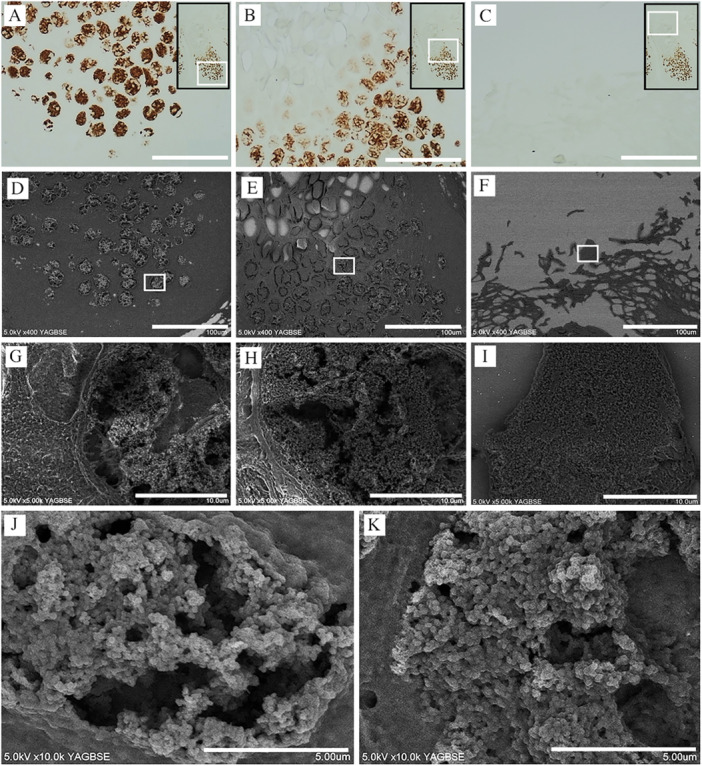
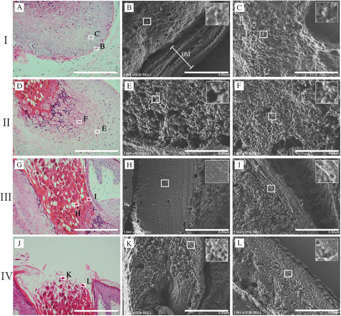
We applied NanoSuit-correlative light and electron microscopy (NanoSuit-CLEM) to formalin-fixed paraffin-embedded (FFPE) skin sections to visualize MC virus particles in situ with high resolution. Melan-A immunohistochemistry using 3,3'-diaminobenzidine (DAB) with osmium staining was performed to identify Henderson-Patterson bodies.
Ultrastructural analysis revealed that MC virus particles were densely localized in the stratum corneum but did not invade deeper epithelial layers in intact skin. However, in areas of epidermal disruption, such as detached or damaged stratum corneum, the virus was observed penetrating into lower layers. While Melan-A immunostaining successfully detected Henderson-Patterson bodies, it failed to identify mature MC virus particles. In contrast, NanoSuit-CLEM combined with Mayer's hematoxylin and lead staining enabled detailed visualization of mature viral particles and their distribution within the stratum corneum.
These findings provide direct ultrastructural evidence that MC virus entry occurs through compromised skin, underscoring the crucial role of the stratum corneum in barrier function. This study highlights the importance of preventing mechanical skin injury, such as scratching or shaving, to limit MC transmission. NanoSuit-CLEM offers a powerful new tool for studying viral pathogenesis in archival tissue samples.
传染性软疣(MC)是由痘病毒科成员引起的常见病毒性皮肤感染。它主要影响儿童、性活跃的成年人和免疫功能低下的个体。尽管MC通过直接接触和自体接种传播,但病毒穿透皮肤屏障的确切机制仍知之甚少。
我们将纳米套装相关光镜和电镜(NanoSuit-CLEM)应用于福尔马林固定石蜡包埋(FFPE)皮肤切片,以高分辨率原位观察MC病毒颗粒。使用3,3'-二氨基联苯胺(DAB)和锇染色进行Melan-A免疫组织化学,以识别亨德森-帕特森小体。
超微结构分析显示,MC病毒颗粒密集地定位于角质层,但在完整皮肤中不侵入更深的上皮层。然而,在表皮破坏区域,如分离或受损的角质层,观察到病毒穿透到下层。虽然Melan-A免疫染色成功检测到亨德森-帕特森小体,但未能识别成熟的MC病毒颗粒。相比之下,NanoSuit-CLEM与 Mayer苏木精和铅染色相结合,能够详细观察成熟病毒颗粒及其在角质层内的分布。
这些发现提供了直接的超微结构证据,表明MC病毒通过受损皮肤进入,强调了角质层在屏障功能中的关键作用。本研究强调了预防机械性皮肤损伤(如抓挠或刮胡子)以限制MC传播的重要性。NanoSuit-CLEM为研究存档组织样本中的病毒发病机制提供了一种强大的新工具。