Institute for NanoSuit Research, Preeminent Medical Photonics Education & Research Center, Hamamatsu University School of Medicine, Hamamatsu, Japan.
Department of Obstetrics and Gynecology, Hamamatsu University School of Medicine, Hamamatsu, Japan.
Lab Invest. 2020 Jan;100(1):161-173. doi: 10.1038/s41374-019-0309-7. Epub 2019 Aug 29.
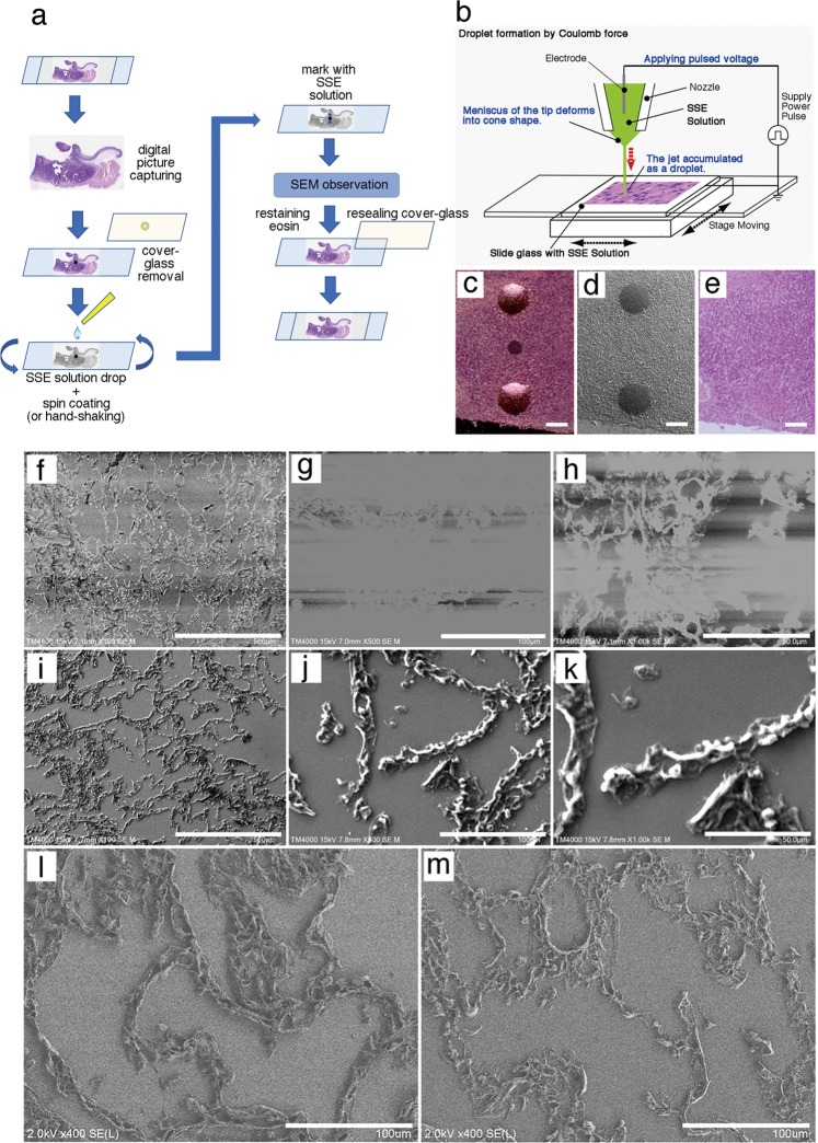
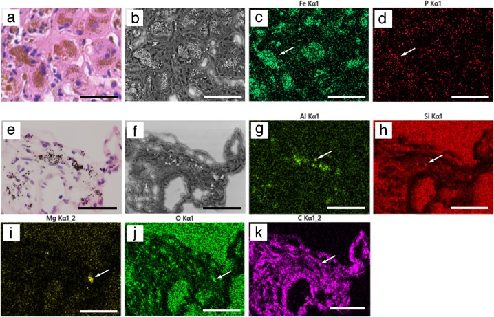
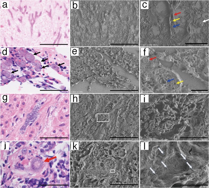
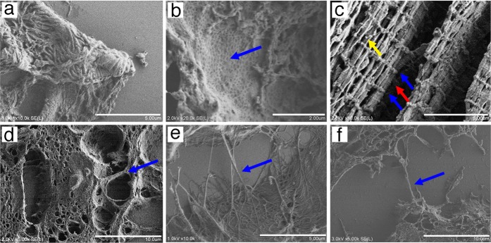
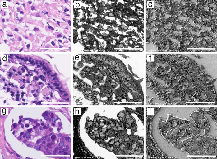
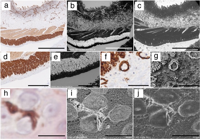
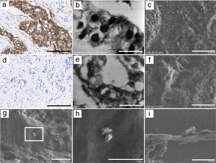
Histological examination using the light microscopy is currently the gold standard for life science research and diagnostics. However, magnified observations are limited because of the limitations intrinsic to light microscopy. Thus, a dual approach, known as correlative light and electron microscopy (CLEM), has emerged, although several technical challenges remain in terms of observing myriad stored paraffin sections. Previously, we developed the NanoSuit method, which enabled us to keep multicellular organisms alive/wet in the high vacuum of a scanning electron microscope by encasing the sample in a thin, vacuum-proof membrane. The approach uses the native extracellular substance (ECS) or an ECS-mimicking substance to polymerize a membrane by plasma or electron beam irradiation. Since the resulting NanoSuit is flexible and dense enough to prevent a living organism's bodily gas and liquids from evaporating (which we refer to as the "surface shield enhancer" (SSE) effect), it works like a miniature spacesuit with sufficient electron conductivity for an SEM observation. Here, we apply the NanoSuit method to CLEM analysis of paraffin sections. Accordingly, the NanoSuit method permits the study of paraffin sections using CLEM at low and high magnification, with the following features: (i) the integrity of the glass slide is maintained, (ii) three-dimensional microstructures of tissue and pathogens are visualized, (iii) nuclei and 3,3'-diaminobenzidine-stained areas are distinct because of gold chloride usage, (iv) immunohistochemical staining is quantitative, and (v) contained elements can be analyzed. Removal of the SSE solution after observation is a further advantage, as this allows slides to be restained and stored. Thus, the NanoSuit method represents a novel approach that will advance the field of histology.
组织学检查使用光学显微镜目前是生命科学研究和诊断的金标准。然而,由于光显微镜的固有局限性,放大观察受到限制。因此,出现了一种称为相关光电子显微镜(CLEM)的双重方法,尽管在观察大量保存的石蜡切片方面仍然存在一些技术挑战。以前,我们开发了 NanoSuit 方法,该方法通过将样品封装在薄的、防真空的膜中,使多细胞生物在扫描电子显微镜的高真空下保持存活/湿润。该方法使用天然细胞外基质(ECS)或 ECS 模拟物质通过等离子体或电子束辐照聚合膜。由于所得的 NanoSuit 足够柔韧和致密,可以防止生物体的气体和液体蒸发(我们称之为“表面屏蔽增强剂”(SSE)效应),因此它就像一个微型太空服一样,具有足够的电子导电性,可用于 SEM 观察。在这里,我们将 NanoSuit 方法应用于石蜡切片的 CLEM 分析。因此,NanoSuit 方法允许在低倍和高倍放大倍率下使用 CLEM 研究石蜡切片,具有以下特点:(i)保持载玻片的完整性,(ii)可视化组织和病原体的三维微观结构,(iii)由于使用氯化金,细胞核和 3,3'-二氨基联苯胺染色区域清晰可见,(iv)免疫组织化学染色是定量的,(v)包含的元素可以被分析。观察后去除 SSE 溶液是另一个优点,因为这允许重新染色和存储载玻片。因此,NanoSuit 方法代表了一种将推进组织学领域的新方法。