He Zhibing, Yang Chao, Zhou Ling, Li Shuang
Department of Nuclear Medicine, Xiangyang No. 1 People's Hospital, Hubei University of Medicine, 15 Jiefang Road, Fan District, Xiangyang, 441000, Hubei, China.
Department of Radiology, Xiangyang No. 1 People's Hospital, Hubei University of Medicine, Xiangyang, China.
Sci Rep. 2025 Jun 6;15(1):20029. doi: 10.1038/s41598-025-02672-x.
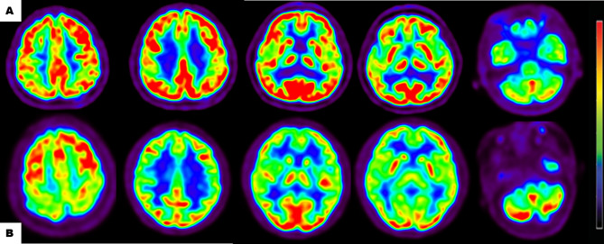
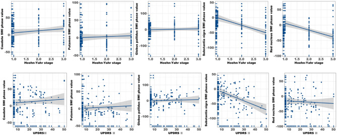
This study aimed to evaluate the diagnostic potential of combining F-FDG PET and susceptibility-weighted imaging (SWI) to assess cerebral glucose metabolism and iron deposition patterns in Parkinson's disease (PD), and to determine their correlations with clinical progression and diagnostic accuracy. Forty-nine PD patients and 70 age-/sex-matched healthy controls underwent standardized F-FDG PET and SWI. Metabolic activity (SUVR) and SWI phase values were quantified in cortical/subcortical regions. Statistical analyses included Mann-Whitney U tests, Pearson/Spearman correlations, and ROC curve analysis to evaluate biomarker-clinical relationships and diagnostic performance. PD patients exhibited hypometabolism in frontal, parietal, and temporal cortices (P < 0.05) and hypermetabolism in the putamen, globus pallidus, and cerebellum (P < 0.05). Cortical hypometabolism correlated with Hoehn-Yahr (H-Y) stages (e.g., temporal lobe: r = - 0.405, P = 0.004) and UPDRS III scores (e.g., frontal cortex: r = - 0.364, P = 0.011). SWI revealed reduced phase values in the substantia nigra, red nucleus, and basal ganglia (P < 0.001), with substantia nigra phase values strongly correlating with H-Y stages (r = - 0.525) and UPDRS III scores (r = - 0.446). Multimodal integration of F-FDG PET and SWI achieved superior diagnostic accuracy (AUC = 0.844) compared to single-modality models (PET: AUC = 0.777; SWI: AUC = 0.780, P < 0.0001). The integration of F-FDG PET and SWI enhances PD diagnosis by capturing complementary metabolic and iron deposition biomarkers. Cortical hypometabolism may precede subcortical iron accumulation, aligning with Braak staging theory. Limitations include cross-sectional design and technical constraints in SWI quantification. Future studies should validate these findings with longitudinal cohorts and advanced techniques like QSM.
本研究旨在评估¹⁸F-氟代脱氧葡萄糖正电子发射断层扫描(¹⁸F-FDG PET)与磁敏感加权成像(SWI)相结合在评估帕金森病(PD)脑葡萄糖代谢和铁沉积模式方面的诊断潜力,并确定它们与临床进展和诊断准确性的相关性。49例PD患者和70例年龄/性别匹配的健康对照者接受了标准化的¹⁸F-FDG PET和SWI检查。对皮质/皮质下区域的代谢活性(标准化摄取值,SUVR)和SWI相位值进行量化。统计分析包括曼-惠特尼U检验、Pearson/Spearman相关性分析和ROC曲线分析,以评估生物标志物与临床的关系及诊断性能。PD患者额叶、顶叶和颞叶皮质表现为代谢减低(P<0.05),壳核、苍白球和小脑表现为代谢增高(P<0.05)。皮质代谢减低与Hoehn-Yahr(H-Y)分期相关(如颞叶:r = -0.405,P = 0.004)和统一帕金森病评定量表第三部分(UPDRS III)评分相关(如额叶皮质:r = -0.364,P = 0.011)。SWI显示黑质、红核和基底神经节的相位值降低(P<0.001),黑质相位值与H-Y分期(r = -0.525)和UPDRS III评分(r = -0.446)密切相关。与单模态模型(PET:AUC = 0.777;SWI:AUC = 0.780,P<0.0001)相比,¹⁸F-FDG PET和SWI的多模态整合实现了更高的诊断准确性(AUC = 0.844)。¹⁸F-FDG PET和SWI的整合通过获取互补的代谢和铁沉积生物标志物提高了PD的诊断水平。皮质代谢减低可能先于皮质下铁蓄积,这与Braak分期理论一致。局限性包括横断面设计和SWI量化中的技术限制。未来的研究应使用纵向队列和如定量磁敏感成像(QSM)等先进技术验证这些发现。