Samarla Suresh Kumar, P Maragathavalli
IT, Puducherry Technological University, Puducherry, India.
CSE, SRKR Engineering college, Bhimavaram, Andhra Pradesh, India.
MethodsX. 2025 May 14;14:103348. doi: 10.1016/j.mex.2025.103348. eCollection 2025 Jun.
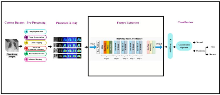
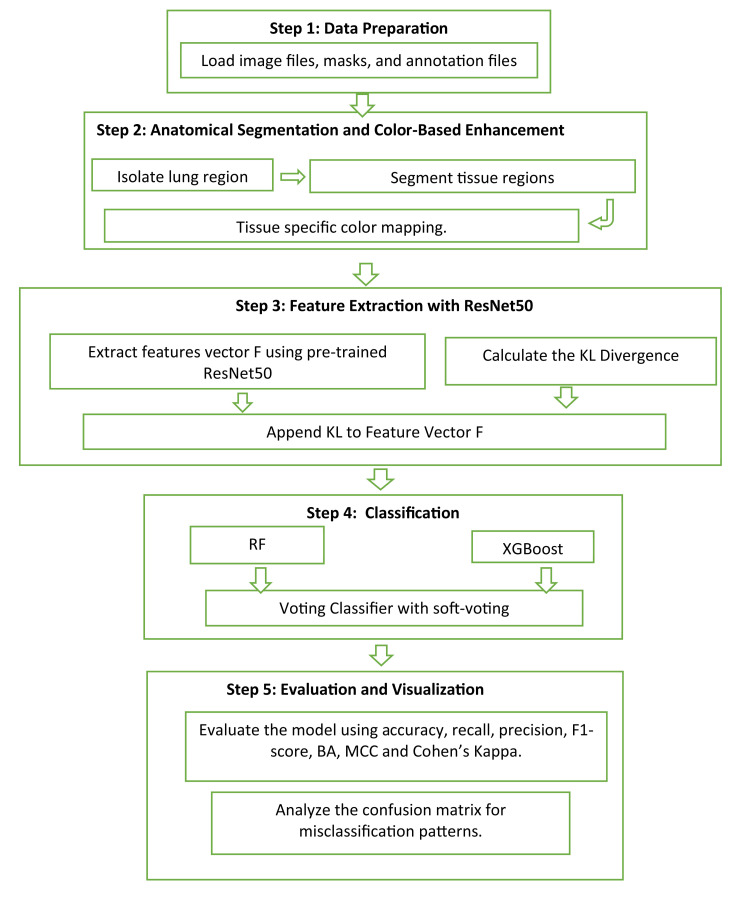
Detecting lung abnormalities via chest X-rays is challenging due to understated tissue variations often ignored by traditional methods. Augmentation techniques like rotation or flipping risk distorting critical anatomical features, actually leading to misdiagnosis. This paper proposes a novel two-stage ASCE (Anatomical Segmentation and Color-Based Enhancement) framework for precise and efficient classification of lung abnormalities while preserving anatomical integrity. Stage 1 classifies Normal vs. Pneumonia with 95 % accuracy, an AUC of 0.98, and an F1-score of 0.92. Stage 2 distinguishes Pneumonia into Viral and Bacterial subtypes with 100 % accuracy and F1-score. This approach integrates segmentation and tissue-specific color enhancements with Kullback-Leibler (KL) divergence, quantifying deviations from healthy lung regions for improved classification. The lightweight pipeline ensures computational efficiency (∼0.06s/image) and clinical interpretability by preserving diagnostic features, enhancing visibility, and enabling quantitative analysis.1. The methodology ensures that diagnostic features are preserved and highlighted with Anatomy-Preserved Segmentation2. The system employs targeted colour-based enhancement that improves the visibility of potential abnormalities3. The model enhances precise identification of abnormal tissue by comparing the probability distributions of healthy lungs and abnormal areas.
通过胸部X光检测肺部异常具有挑战性,因为传统方法常常忽略了细微的组织差异。诸如旋转或翻转等增强技术有扭曲关键解剖特征的风险,实际上会导致误诊。本文提出了一种新颖的两阶段ASCE(解剖分割和基于颜色的增强)框架,用于在保持解剖完整性的同时精确高效地对肺部异常进行分类。第一阶段对正常与肺炎进行分类,准确率为95%,AUC为0.98,F1分数为0.92。第二阶段将肺炎区分为病毒和细菌亚型,准确率和F1分数均为100%。该方法将分割和组织特异性颜色增强与库尔贝克-莱布勒(KL)散度相结合,量化与健康肺区域的偏差以改进分类。轻量级流程通过保留诊断特征、提高可见性和实现定量分析来确保计算效率(约0.06秒/图像)和临床可解释性。1. 该方法通过解剖保留分割确保诊断特征得以保留并突出显示。2. 该系统采用有针对性的基于颜色的增强,提高潜在异常的可见性。3. 该模型通过比较健康肺和异常区域的概率分布来增强对异常组织的精确识别。