Fathimah Fariztah Sukainah Nur, Ari Widjaja Sauli, Sasono Wimbo, Yustiarini Ima, Firmansjah Muhammad, Prakosa Ady Dwi, Mulyazhara Aulia Kezia, Soelistijo Soebagijo Adi
Department of Ophthalmology, Faculty of Medicine, Universitas Airlangga, Surabaya, East Java, Indonesia.
Department of Ophthalmology, Dr. Soetomo General Academic Hospital, Surabaya, East Java, Indonesia.
Int J Retina Vitreous. 2025 Jun 12;11(1):64. doi: 10.1186/s40942-025-00688-z.
Retinal vessel geometry characteristic have been studied as one of the signs of microvascular changes in diabetic retinopathy (DR) that necessitates early screening. This study aimed to investigate the differences in retinal vessel tortuosity (VT) and fractal dimension (FD) between patients with and without DR.
This retrospective study analyzed medical records and OCT-A images of DR and No-DR patients. DR severity was graded by a vitreoretinal specialist following the International Clinical Diabetic Retinopathy and Diabetic Macular Edema Severity Scales. Retinal VT and FD were quantified using ImageJ software. Comparison between groups using non-parametric and Generalized Estimating Equations (GEE) statistical analysis combined with cluster bootstrapping.
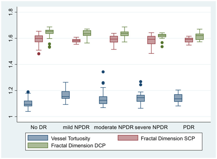
We analyzed 96 (161 eyes) with the mean age of 52.7 ± 9.9 years. Compared to No-DR, VT was significantly higher in all DR groups (p < 0.05). Mild non proliferative DR (β = +0.0621), Moderate NPDR (β = +0.0412), Severe NPDR (β = +0.0441), and proliferative DR (β = +0.0404). FD of the superficial capillary plexus (SCP) showed no significant difference among the groups and a significantly lower FD of the deep capillary plexus (DCP) compared to the No-DR groups (moderate NPDR (β = -0.0131), severe NPDR ( β = -0.0316) and PDR ( β = -0.0326)).
Compared to No-DR group, VT was found significantly higher in DR group, and FD of the DCP found significantly lower in the DR group. These parameters offer unique insights beyond simple vessel loss and complementary information into the geometric complexity and structural alterations of the retinal microvasculature in DR.
视网膜血管几何特征已被作为糖尿病视网膜病变(DR)微血管变化的标志之一进行研究,这需要早期筛查。本研究旨在调查糖尿病视网膜病变患者与非糖尿病视网膜病变患者之间视网膜血管迂曲度(VT)和分形维数(FD)的差异。
这项回顾性研究分析了糖尿病视网膜病变患者和非糖尿病视网膜病变患者的病历及光学相干断层扫描血管造影(OCT-A)图像。糖尿病视网膜病变的严重程度由玻璃体视网膜专科医生根据国际临床糖尿病视网膜病变和糖尿病黄斑水肿严重程度量表进行分级。使用ImageJ软件对视网膜VT和FD进行量化。采用非参数和广义估计方程(GEE)统计分析并结合聚类自抽样进行组间比较。
我们分析了96例患者(161只眼),平均年龄为52.7±9.9岁。与非糖尿病视网膜病变患者相比,所有糖尿病视网膜病变组的VT均显著更高(p<0.05)。轻度非增殖性糖尿病视网膜病变(β = +0.0621)、中度非增殖性糖尿病视网膜病变(β = +0.0412)、重度非增殖性糖尿病视网膜病变(β = +0.0441)和增殖性糖尿病视网膜病变(β = +0.0404)。浅表毛细血管丛(SCP)的FD在各组之间无显著差异,与非糖尿病视网膜病变组相比,深层毛细血管丛(DCP)的FD显著更低(中度非增殖性糖尿病视网膜病变(β = -0.0131)、重度非增殖性糖尿病视网膜病变(β = -0.0316)和增殖性糖尿病视网膜病变(β = -0.0326))。
与非糖尿病视网膜病变组相比,糖尿病视网膜病变组的VT显著更高,糖尿病视网膜病变组的DCP的FD显著更低。这些参数除了能提供简单的血管丢失之外,还能对糖尿病视网膜病变中视网膜微血管的几何复杂性和结构改变提供独特见解及补充信息。