Alosaimi Abdulrahman, Hafiz Badr E, Tawfiq Ibrahim A, Makhdoom Naeem, Almoghthwey Talal H
Otolaryngology, Head and Neck Surgery, Ohud Hospital, Medinah, SAU.
Neurological Surgery, King Faisal Specialist Hospital and Research Centre, Jeddah, SAU.
Cureus. 2025 May 24;17(5):e84721. doi: 10.7759/cureus.84721. eCollection 2025 May.
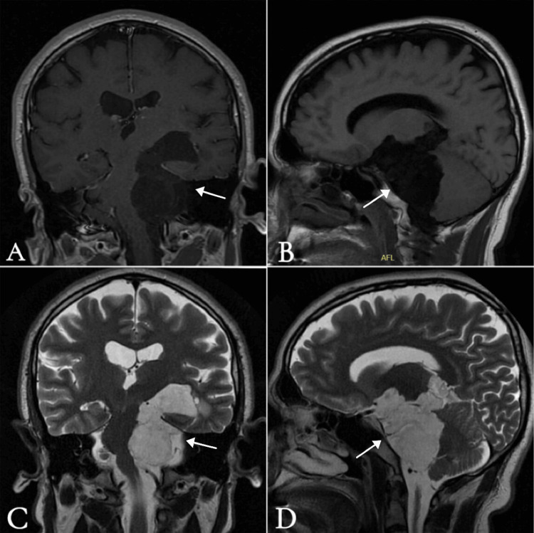
Epidermoid cysts are rare congenital tumors of the central nervous system. These histologically benign, slow-growing lesions form when ectodermal cells become trapped during the closure of the neural tube. Histologically, they consist of a core composed of keratin, desquamated epithelial cells, and cholesterol, surrounded by a layer of stratified squamous epithelium. Clinical features depend on the lesion's location. In the cerebellopontine angle (CPA), they typically present with tinnitus, vertigo, hearing loss, and facial weakness, with or without cerebellar signs and symptoms. Unilateral hearing loss as the sole presenting symptom is uncommon in the setting of a large, extensive cyst and may delay diagnosis. A 35-year-old male presented with progressive left-sided hearing loss for one year, without vertigo, tinnitus, or other neurological symptoms. Audiological testing revealed severe-to-profound sensorineural hearing loss in the left ear. Temporal bone computed tomography and brain magnetic resonance imaging showed a large, extra-axial cystic lesion in the left CPA with characteristic diffusion-weighted imaging restriction, consistent with an epidermoid cyst. The lesion caused significant mass effect, including compression of the brainstem, cranial nerves, basilar artery, left vertebral artery, and left posterior cerebral artery. The patient underwent successful surgical excision via a retrosigmoid suboccipital craniotomy. Histopathological examination confirmed the diagnosis of an epidermoid cyst. This case report highlights an unusual presentation of an extensive epidermoid cyst in the left CPA that manifested solely as unilateral hearing loss, underscoring the diagnostic challenges posed by this rare lesion. The findings emphasize the importance of considering atypical presentations of intracranial tumors in the differential diagnosis of patients with unexplained hearing loss.
表皮样囊肿是中枢神经系统罕见的先天性肿瘤。这些组织学上良性、生长缓慢的病变是在神经管闭合期间外胚层细胞被困住时形成的。组织学上,它们由一个核心组成,该核心由角蛋白、脱落的上皮细胞和胆固醇构成,周围是一层复层鳞状上皮。临床特征取决于病变的位置。在桥小脑角(CPA),它们通常表现为耳鸣、眩晕、听力丧失和面部无力,伴有或不伴有小脑体征和症状。在巨大、广泛的囊肿情况下,单侧听力丧失作为唯一的表现症状并不常见,可能会延迟诊断。一名35岁男性出现进行性左侧听力丧失一年,无眩晕、耳鸣或其他神经症状。听力测试显示左耳有重度至极重度感音神经性听力丧失。颞骨计算机断层扫描和脑部磁共振成像显示左侧CPA有一个巨大的轴外囊性病变,具有特征性的扩散加权成像受限,符合表皮样囊肿。该病变引起明显的占位效应,包括压迫脑干、颅神经、基底动脉、左侧椎动脉和左侧大脑后动脉。患者通过乙状窦后枕下开颅手术成功切除病变。组织病理学检查证实为表皮样囊肿。本病例报告强调了左侧CPA广泛表皮样囊肿的一种不寻常表现,仅表现为单侧听力丧失,突出了这种罕见病变带来的诊断挑战。这些发现强调了在不明原因听力丧失患者的鉴别诊断中考虑颅内肿瘤非典型表现的重要性。