Ghasemi Iman, Gogiraju Rajinikanth, Khraisat Sana'a, Pagel Sven, Graf Claudine, Brandt Moritz, Madhusudhan Thati, Wenzel Philip, Luxán Guillermo, Lurz Philipp, Bochenek Magdalena L, Schäfer Katrin
Department of Cardiology, Cardiology I, University Medical Center of the Johannes Gutenberg University, D-55131 Mainz, Germany.
Center for Thrombosis and Hemostasis, University Medical Center of the Johannes Gutenberg University, D-55131 Mainz, Germany.
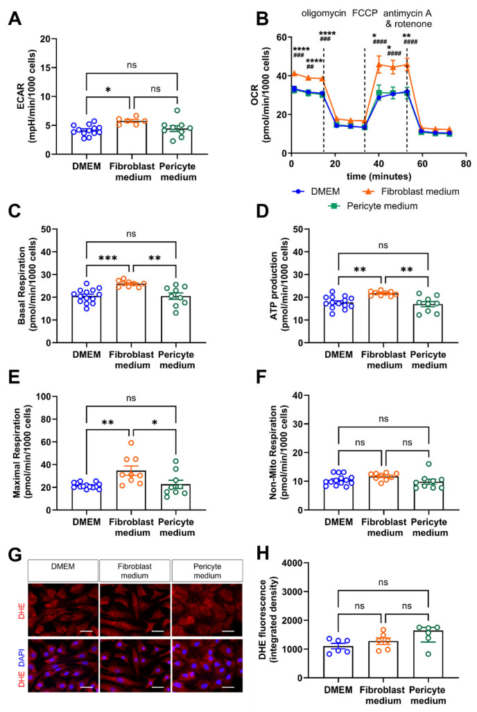
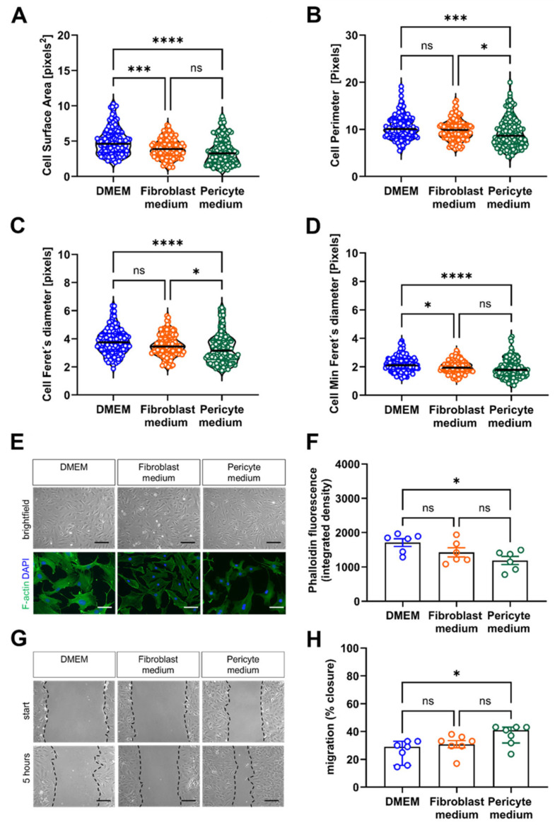
Cells. 2025 Jun 19;14(12):927. doi: 10.3390/cells14120927.
(1) Background: Vascular mural cells reside in the media and outer layers of the vessel wall. Their ability to proliferate and migrate or to change phenotype in response to external cues is a central feature of the vascular response to injury. Genetically engineered mice are used for loss- or gain-of-function analyses or lineage tracing in vivo, their primary cells for mechanistic studies in vitro. Whether and how cultivation conditions affect their phenotype and function is often overlooked. (2) Methods: Here, we systematically studied how the cultivation of primary mural cells isolated from the aorta of adult wild-type mice in either basal medium (DMEM) or special media formulated for the cultivation of fibroblasts or pericytes affects their phenotype and function. (3) Results: Medium composition did not alter cell viability, but the mRNA levels of differentiated smooth muscle cell markers were highest in vascular mural cells expanded in DMEM. Conversely, significantly higher numbers of proliferating and migrating cells were observed in cells expanded in Pericyte medium, and cytoskeletal rearrangements supported increased migratory capacities. Significantly reduced telomere lengths and metabolic reprogramming was observed in aortic mural cells cultured in Fibroblast medium. (4) Conclusions: Our findings underline the plasticity of primary aortic mural cells and highlight the importance of the culture media composition during their expansion, which could be exploited to interrogate their responsiveness to external stimuli or conditions observed in vivo or in patients.
(1) 背景:血管壁细胞位于血管壁的中膜和外层。它们响应外部信号进行增殖、迁移或改变表型的能力是血管对损伤反应的核心特征。基因工程小鼠用于体内功能缺失或获得分析或谱系追踪,其原代细胞用于体外机制研究。培养条件是否以及如何影响它们的表型和功能常常被忽视。(2) 方法:在此,我们系统地研究了将从成年野生型小鼠主动脉分离的原代壁细胞在基础培养基(DMEM)或为培养成纤维细胞或周细胞配制的特殊培养基中培养如何影响它们的表型和功能。(3) 结果:培养基成分未改变细胞活力,但在DMEM中扩增的血管壁细胞中,分化的平滑肌细胞标志物的mRNA水平最高。相反,在周细胞培养基中扩增的细胞中观察到增殖和迁移细胞的数量显著增加,细胞骨架重排支持迁移能力增强。在成纤维细胞培养基中培养的主动脉壁细胞中观察到端粒长度显著缩短和代谢重编程。(4) 结论:我们的研究结果强调了原代主动脉壁细胞的可塑性,并突出了其扩增过程中培养基成分的重要性,这可用于研究它们对体内或患者中观察到的外部刺激或条件的反应性。