Moazamian Dina, Daskareh Mahyar, Athertya Jiyo S, Suprana Arya A, Jerban Saeed, Ma Yajun
Department of Radiology, University of California, San Diego, CA 92037, USA.
Department of Bioengineering, University of California, San Diego, CA 92037, USA.
J Imaging. 2025 Jun 16;11(6):198. doi: 10.3390/jimaging11060198.
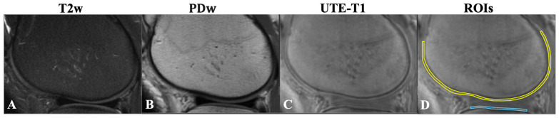
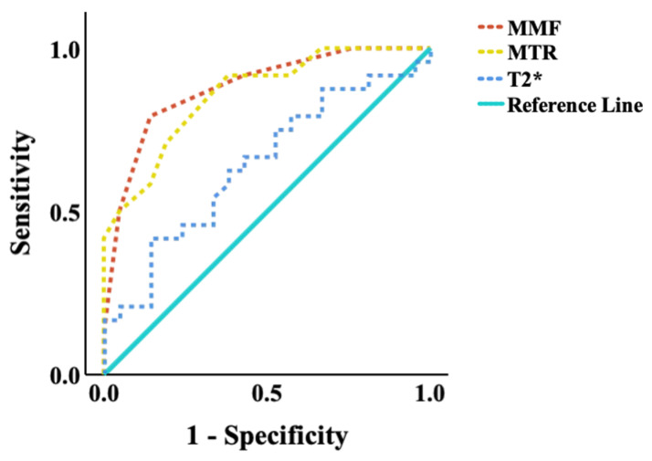
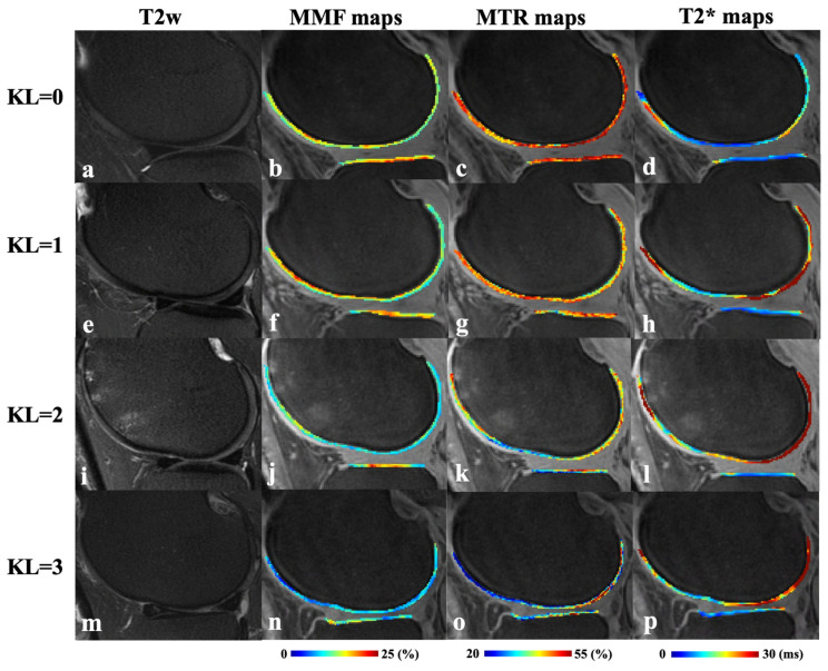
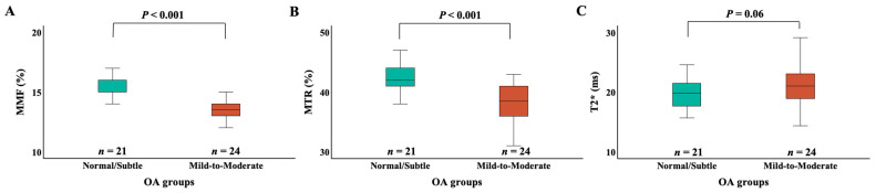
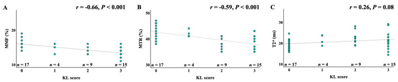
Osteoarthritis (OA) is the most prevalent degenerative joint disorder worldwide, causing significant declines in quality of life. The osteochondral junction (OCJ), a critical structural interface between deep cartilage and subchondral bone, plays an essential role in OA progression but is challenging to assess using conventional magnetic resonance imaging (MRI) due to its short T2 relaxation times. This study aimed to evaluate the utility of ultrashort echo time (UTE) MRI biomarkers, including macromolecular fraction (MMF), magnetization transfer ratio (MTR), and T2*, for in vivo quantification of OCJ changes in knee OA for the first time. Forty-five patients (mean age: 53.8 ± 17.0 years, 50% female) were imaged using 3D UTE-MRI sequences on a 3T clinical MRI scanner. Patients were stratified into two OA groups based on radiographic Kellgren-Lawrence (KL) scores: normal/subtle (KL = 0-1) ( = 21) and mild to moderate (KL = 2-3) ( = 24). Quantitative analysis revealed significantly lower MMF (15.8 ± 1.4% vs. 13.6 ± 1.2%, < 0.001) and MTR (42.5 ± 2.5% vs. 38.2 ± 2.3%, < 0.001) in the higher KL 2-3 group, alongside a higher trend in T2* values (19.7 ± 2.6 ms vs. 21.6 ± 3.8 ms, = 0.06). Moreover, MMF and MTR were significantly negatively correlated with KL grades (r = -0.66 and -0.59; < 0.001, respectively), while T2* showed a weaker positive correlation (r = 0.26, = 0.08). Receiver operating characteristic (ROC) analysis demonstrated superior diagnostic accuracy for MMF (AUC = 0.88) and MTR (AUC = 0.86) compared to T2* (AUC = 0.64). These findings highlight UTE-MT techniques (i.e., MMF and MTR) as promising imaging tools for detecting OCJ degeneration in knee OA, with potential implications for earlier and more accurate diagnosis and disease monitoring.
骨关节炎(OA)是全球最常见的退行性关节疾病,会导致生活质量显著下降。骨软骨交界处(OCJ)是深层软骨与软骨下骨之间的关键结构界面,在OA进展中起重要作用,但由于其T2弛豫时间短,使用传统磁共振成像(MRI)进行评估具有挑战性。本研究旨在首次评估超短回波时间(UTE)MRI生物标志物,包括大分子分数(MMF)、磁化传递率(MTR)和T2*,用于体内定量评估膝关节OA中OCJ变化的效用。45例患者(平均年龄:53.8±17.0岁,50%为女性)在3T临床MRI扫描仪上使用3D UTE-MRI序列进行成像。根据放射学Kellgren-Lawrence(KL)评分将患者分为两个OA组:正常/轻微(KL=0-1)(n=21)和轻度至中度(KL=2-3)(n=24)。定量分析显示,KL 2-3较高组的MMF(15.8±1.4%对13.6±1.2%,P<0.001)和MTR(42.5±2.5%对38.2±2.3%,P<0.001)显著降低,同时T2值有升高趋势(19.7±2.6毫秒对21.6±3.8毫秒,P=0.06)。此外,MMF和MTR与KL分级显著负相关(r=-0.66和-0.59;P分别<0.001),而T2显示出较弱的正相关(r=0.26,P=0.08)。受试者操作特征(ROC)分析表明,与T2*(AUC=0.64)相比,MMF(AUC=0.88)和MTR(AUC=0.86)具有更高的诊断准确性。这些发现突出了UTE-MT技术(即MMF和MTR)作为检测膝关节OA中OCJ退变的有前景的成像工具,对早期和更准确的诊断及疾病监测具有潜在意义。