Bayeh Betsega A, Marchand-Adam Sylvain, Legué Sylvie, Luque Paz David, Plantier Laurent, Flament Thomas
Tours University Hospital, Centre de Référence des Maladies Pulmonaires Rares de la Région Centre-Val-de-Loire, Hôpital Bretonneau, 37000 Tours, France.
Faculté de Médecine, Tours University, Centre d'Étude des Pathologies Respiratoires (CEPR) INSERM U1100, 37032 Tours, France.
J Clin Med. 2025 Jun 11;14(12):4159. doi: 10.3390/jcm14124159.
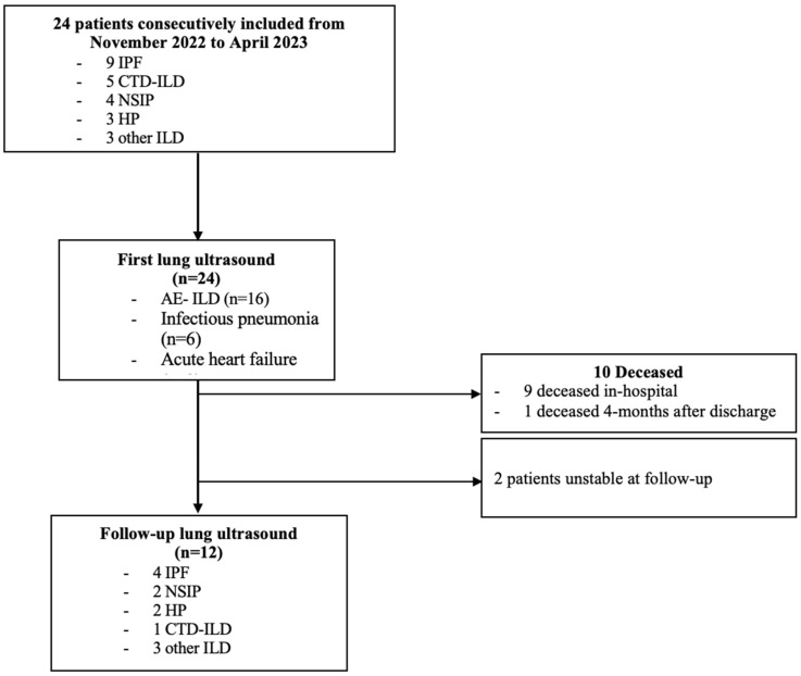
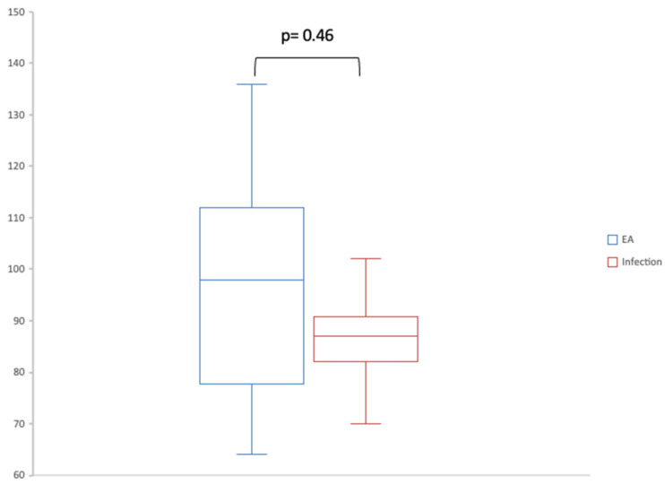
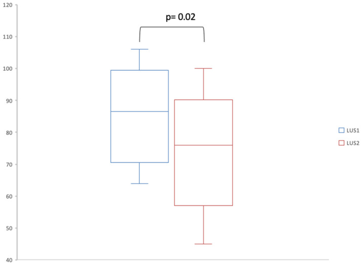
Lung ultrasound (LUS) can be used at follow-up for patients with stable interstitial lung disease (ILD). LUS could also help guide the diagnosis of etiology for acute respiratory episodes. We conducted a prospective, one-center, observational study including patients with ILD hospitalized in the pulmonology unit or in the intensive care unit of the Tours University Hospital for acute dyspnea. LUS was performed at admission and then at a follow-up visit in the six months following discharge. We compared the number of B-lines between the two LUSs. We also compared the features of the first LUS between the different etiologies responsible for increased dyspnea. Of 24 patients, 16 had acute ILD exacerbation (67%), 6 had pulmonary infections (25%) and 2 had acute heart failure (8%). LUS was feasible in all patients and always showed lung sliding, pleural irregularities and B-lines. There were pleural effusions in four cases (17%) and pulmonary consolidations in two cases (8%). Seven patients had A-lines in at least one thoracic space on the initial LUS. We found a significant decrease in the number of B-lines at follow-up (76; IQR, [59-86.75]) compared to admission (86.5; IQR, [71.5-94.5]) (-value = 0.02). There was a trend of more A-lines in patients with infection (1 [0.25-1.75]) compared to AE-ILD (0 [0-0]). Following an episode of acute dyspnea in patients with ILD, LUS shows a decrease in the number of B-lines. Patients with ILD and concurrent pulmonary infection may have more A-lines than patients with AE-ILD.
肺部超声(LUS)可用于稳定期间质性肺疾病(ILD)患者的随访。LUS还可有助于指导急性呼吸发作病因的诊断。我们进行了一项前瞻性、单中心观察性研究,纳入了因急性呼吸困难入住图尔大学医院肺病科或重症监护病房的ILD患者。在入院时以及出院后6个月的随访时进行LUS检查。我们比较了两次LUS检查时B线的数量。我们还比较了导致呼吸困难加重的不同病因患者首次LUS检查的特征。24例患者中,16例为急性ILD加重(67%),6例为肺部感染(25%),2例为急性心力衰竭(8%)。LUS在所有患者中均可行,且总是显示肺滑动、胸膜不规则和B线。4例(17%)有胸腔积液,2例(8%)有肺实变。7例患者在初次LUS检查时至少一个胸腔有A线。我们发现随访时B线数量(76;四分位数间距,[59 - 86.75])较入院时(86.5;四分位数间距,[71.5 - 94.5])显著减少(P值 = 0.02)。与急性加重期ILD患者(0 [0 - 0])相比,感染患者(1 [0.25 - 1.75])的A线有增多趋势。ILD患者发生急性呼吸困难发作后,LUS显示B线数量减少。与急性加重期ILD患者相比,合并肺部感染的ILD患者可能有更多A线。