Boylan Brendan T, Hwang Mihyun, Brozost Elyse, Oh Hyunsuk, Tumanov Alexei V, Louveau Antoine, Bergmann Cornelia C
Department of Neurosciences, Lerner Research Institute, Cleveland Clinic Foundation, Cleveland, OH, 44195, USA.
Department of Pathology, Case Western Reserve University School of Medicine, Cleveland, OH, 44106, USA.
J Neuroinflammation. 2025 Jun 27;22(1):165. doi: 10.1186/s12974-025-03491-7.
CNS stromal cells, especially fibroblasts and endothelial cells, support leukocyte accumulation through upregulation of adhesion molecules and lymphoid chemokines. While chronically activated fibroblast networks can drive pathogenic immune cell aggregates known as tertiary lymphoid structures (TLS), early stromal cell activation during CNS infection can support anti-viral T cells. However, the cell types and factors driving early stromal cell activation is poorly explored.
A neurotropic murine coronavirus (mCoV) infection model was used to better characterize signals that promote fibroblast networks supporting accumulation of antiviral lymphocytes. Based on the early appearance of IgD B cells with unknown functions during several CNS infections, we probed their potential to activate stromal cells through lymphotoxin β (LTβ), a molecule critical in maintaining fibroblast-networks in lymphoid tissues as well as promoting TLS in autoimmunity and cancers.
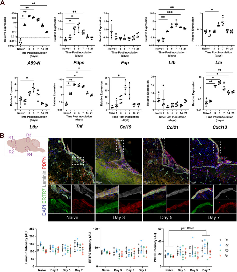
Kinetic analysis of stromal cell activation in olfactory bulbs and brains revealed that upregulation of adhesion molecules and lymphoid chemokines Ccl19, Ccl21 and Cxcl13 closely tracked viral replication. Immunohistochemistry revealed that upregulation of the fibroblast marker podoplanin (PDPN) at meningeal and perivascular sites mirrored kinetics of RNA expression. Moreover, both B cells and T cells colocalized to areas of PDPN reactivity, supporting a potential role in regulating stromal cell activation. However, specific depletion of LTβ from B cells using Mb1-creERT2 x Ltβ mice had no effect on T or B cell recruitment or viral replication. B cell depletion by anti-CD20 antibody also had no adverse effects. Surprisingly, LTβR agonism reduced viral control and parenchymal T cell localization despite increasing stromal cell lymphoid chemokines and PDPN. Additional assessment of direct stromal cell activation by the viral RNA mimic poly I:C showed induction of Pdpn and Ccl19 preceding Ltb.
Neither B cell-derived LTβ or B cells are primary drivers of stromal cell activation networks in the CNS following mCoV infection. Although supplementary agonist mediated LTβR engagement confirmed a role for LTβ in enhancing PDPN and lymphoid chemokine expression, it impeded T cell migration to the CNS parenchyma and viral control. Our data overall indicate that stromal cells can integrate LTβR signals to tune their activation, but that LTβ is not necessarily essential and can even dysregulate protective antiviral T cell functions.
中枢神经系统(CNS)基质细胞,尤其是成纤维细胞和内皮细胞,通过上调黏附分子和淋巴样趋化因子来支持白细胞聚集。虽然长期激活的成纤维细胞网络可驱动称为三级淋巴结构(TLS)的致病性免疫细胞聚集,但中枢神经系统感染期间早期基质细胞的激活可支持抗病毒T细胞。然而,驱动早期基质细胞激活的细胞类型和因素尚未得到充分研究。
使用嗜神经性小鼠冠状病毒(mCoV)感染模型,以更好地表征促进支持抗病毒淋巴细胞聚集的成纤维细胞网络的信号。基于在几种中枢神经系统感染期间功能未知的IgD B细胞的早期出现,我们探究了它们通过淋巴毒素β(LTβ)激活基质细胞的潜力,LTβ是维持淋巴组织中纤维母细胞网络以及在自身免疫和癌症中促进TLS的关键分子。
对嗅球和大脑中基质细胞激活的动力学分析表明,黏附分子和淋巴样趋化因子Ccl19、Ccl21和Cxcl13的上调与病毒复制密切相关。免疫组织化学显示,脑膜和血管周围部位成纤维细胞标志物血小板内皮细胞黏附分子(PDPN)的上调反映了RNA表达的动力学。此外,B细胞和T细胞都共定位于PDPN反应性区域,支持其在调节基质细胞激活中的潜在作用。然而,使用Mb1-creERT2 x Ltβ小鼠特异性去除B细胞中的LTβ对T或B细胞募集或病毒复制没有影响。用抗CD20抗体耗尽B细胞也没有不良影响。令人惊讶的是,尽管增加了基质细胞淋巴样趋化因子和PDPN,但LTβR激动作用降低了病毒控制和实质T细胞定位。对病毒RNA模拟物聚肌苷酸胞苷酸(poly I:C)直接激活基质细胞的进一步评估显示,在Ltb之前诱导了Pdpn和Ccl19。
mCoV感染后,B细胞衍生的LTβ或B细胞都不是中枢神经系统中基质细胞激活网络的主要驱动因素。尽管补充激动剂介导的LTβR参与证实了LTβ在增强PDPN和淋巴样趋化因子表达中的作用,但它阻碍了T细胞向中枢神经系统实质的迁移和病毒控制。我们的数据总体表明,基质细胞可以整合LTβR信号来调节其激活,但LTβ不一定是必需的,甚至可能失调保护性抗病毒T细胞功能。