Roby Merjulah, Restrepo Juan C, Shan Deepak K, Muluk Satish C, Eskandari Mark K, Kashyap Vikram S, Finol Ender A
Department of Mechanical, Aerospace, and Industrial Engineering, The University of Texas at San Antonio, San Antonio TX, 78249, USA.
Department of Thoracic and Cardiovascular Surgery, Allegheny Health Network, Allegheny General Hospital, Pittsburgh PA, 15212, USA.
Res Sq. 2025 Jun 12:rs.3.rs-6630234. doi: 10.21203/rs.3.rs-6630234/v1.
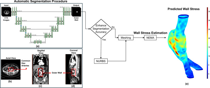
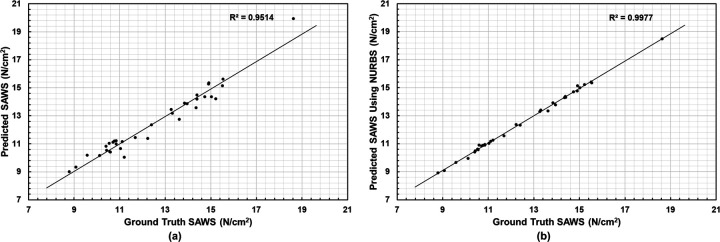
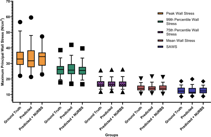
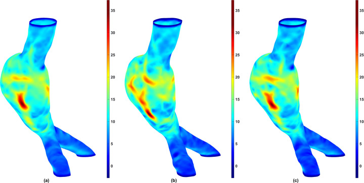
Abdominal Aortic Aneurysm (AAA) remains a significant public health challenge, with an 82.1% increase in related fatalities from 1990 to 2019. In the United States alone, AAA complications resulted in an estimated 13,640 deaths between 2018 and 2021. In clinical practice, computed tomography angiography (CTA) is the primary imaging modality for monitoring and pre-surgical planning of AAA patients. CTA provides high-resolution vascular imaging, enabling detailed assessments of aneurysm morphology and informing critical clinical decisions. However, manual segmentation of CTA images is labor intensive and time consuming, underscoring the need for automated segmentation algorithms, particularly when feature extraction from clinical images can inform treatment decisions. We propose a framework to automatically segment the outer wall of the abdominal aorta from CTA images and estimate AAA wall stress. Our approach employs a patch-based dilated modified U-Net model to accurately delineate the outer wall boundary of AAAs and and Nonlinear Elastic Membrane Analysis (NEMA) to estimate their wall stress. We further integrate Non-Uniform Rational B-Splines (NURBS) to refine the segmentation. During prediction, our deep learning architecture requires 17 ° 0.02 milliseconds per frame to generate the final segmented output. The latter is used to provide critical insight into the biomechanical state of stress of an AAA. This modeling strategy merges advanced deep learning architecture, the precision of NURBS, and the advantages of NEMA to deliver a robust, accurate, and efficient method for computational analysis of AAAs.
腹主动脉瘤(AAA)仍然是一项重大的公共卫生挑战,1990年至2019年期间,相关死亡人数增加了82.1%。仅在美国,2018年至2021年期间,AAA并发症估计导致13640人死亡。在临床实践中,计算机断层扫描血管造影(CTA)是监测AAA患者和术前规划的主要成像方式。CTA提供高分辨率血管成像,能够详细评估动脉瘤形态并为关键临床决策提供依据。然而,CTA图像的手动分割劳动强度大且耗时,这凸显了对自动分割算法的需求,特别是当从临床图像中提取特征可以为治疗决策提供依据时。我们提出了一个框架,用于从CTA图像中自动分割腹主动脉外壁并估计AAA壁应力。我们的方法采用基于补丁的扩张改进U-Net模型来准确描绘AAA的外壁边界,并采用非线性弹性膜分析(NEMA)来估计其壁应力。我们进一步集成非均匀有理B样条(NURBS)来优化分割。在预测过程中,我们的深度学习架构每帧需要17±0.02毫秒来生成最终的分割输出。后者用于深入了解AAA的生物力学应力状态。这种建模策略融合了先进的深度学习架构、NURBS的精度和NEMA的优势,为AAA的计算分析提供了一种强大、准确且高效的方法。