• 文献检索
  • 文档翻译
  • 深度研究
  • 学术资讯
  • Suppr Zotero 插件Zotero 插件
  • 邀请有礼
  • 套餐&价格
  • 历史记录
应用&插件
Suppr Zotero 插件Zotero 插件浏览器插件Mac 客户端Windows 客户端微信小程序
定价
高级版会员购买积分包购买API积分包
服务
文献检索文档翻译深度研究API 文档MCP 服务
关于我们
关于 Suppr公司介绍联系我们用户协议隐私条款
关注我们

Suppr 超能文献

核心技术专利:CN118964589B侵权必究
粤ICP备2023148730 号-1Suppr @ 2026

文献检索

告别复杂PubMed语法,用中文像聊天一样搜索,搜遍4000万医学文献。AI智能推荐,让科研检索更轻松。

立即免费搜索

文件翻译

保留排版,准确专业,支持PDF/Word/PPT等文件格式,支持 12+语言互译。

免费翻译文档

深度研究

AI帮你快速写综述,25分钟生成高质量综述,智能提取关键信息,辅助科研写作。

立即免费体验

深度学习流水线,用于自动进行高分辨率动脉分割,无论是否有静脉对比。

A Deep Learning Pipeline to Automate High-Resolution Arterial Segmentation With or Without Intravenous Contrast.

机构信息

Nuffield Department of Surgical Sciences, University of Oxford, Oxford, United Kingdom.

Department of Engineering Science, University of Oxford, Oxford, United Kingdom.

出版信息

Ann Surg. 2022 Dec 1;276(6):e1017-lpagee1027. doi: 10.1097/SLA.0000000000004595. Epub 2020 Nov 23.

DOI:10.1097/SLA.0000000000004595
PMID:33234786
原文链接:https://pmc.ncbi.nlm.nih.gov/articles/PMC9645535/
Abstract

BACKGROUND

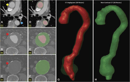
Existing methods to reconstruct vascular structures from a computerized tomography (CT) angiogram rely on contrast injection to enhance the radio-density within the vessel lumen. However, pathological changes in the vasculature may be present that prevent accurate reconstruction. In aortic aneurysmal disease, a thrombus adherent to the aortic wall within the expanding aneurysmal sac is present in >90% of cases. These deformations prevent the automatic extraction of vital clinical information by existing image reconstruction methods.

AIM

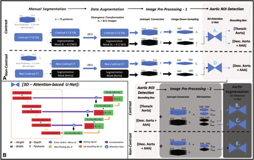
In this study, a deep learning architecture consisting of a modified U-Net with attention-gating was implemented to establish a high-throughput and automated segmentation pipeline of pathological blood vessels in CT images acquired with or without the use of a contrast agent.

METHODS AND RESULTS

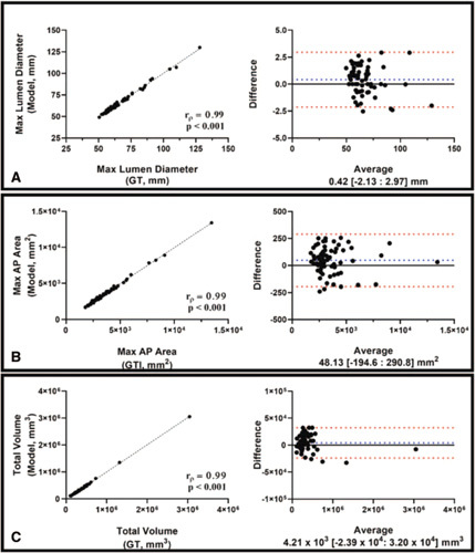
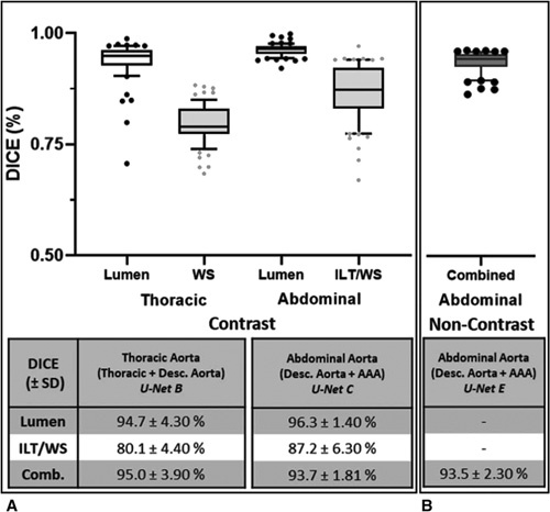
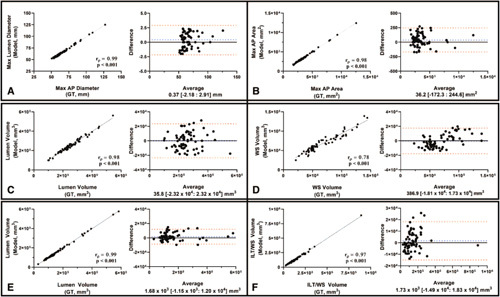
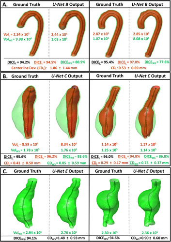
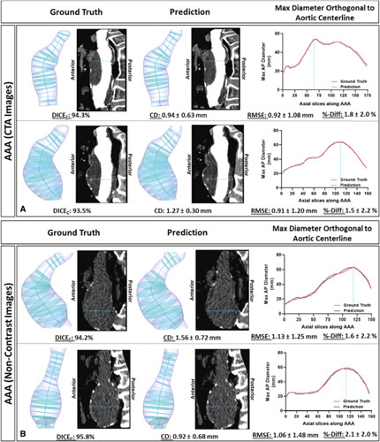
Seventy-Five patients with paired noncontrast and contrast-enhanced CT images were randomly selected from an ongoing study (Ethics Ref 13/SC/0250), manually annotated and used for model training and evaluation. Data augmentation was implemented to diversify the training data set in a ratio of 10:1. The performance of our Attention-based U-Net in extracting both the inner (blood flow) lumen and the wall structure of the aortic aneurysm from CT angiograms was compared against a generic 3-D U-Net and displayed superior results. Implementation of this network within the aortic segmentation pipeline for both contrast and noncontrast CT images has allowed for accurate and efficient extraction of the morphological and pathological features of the entire aortic volume.

CONCLUSIONS

This extraction method can be used to standardize aneurysmal disease management and sets the foundation for complex geometric and morphological analysis. Furthermore, this pipeline can be extended to other vascular pathologies.

摘要

背景

现有的从计算机断层扫描(CT)血管造影重建血管结构的方法依赖于对比剂注射来增强血管腔内的放射密度。然而,可能存在血管的病理性变化,从而导致重建不准确。在主动脉瘤疾病中,在扩张的动脉瘤囊内附着于主动脉壁的血栓在>90%的病例中存在。这些变形阻止了现有图像重建方法自动提取重要的临床信息。

目的

在这项研究中,实现了一个由带有注意力门控的修改后的 U-Net 组成的深度学习架构,以建立一个在使用或不使用造影剂的 CT 图像中对病理性血管进行高通量和自动化分割的流水线。

方法和结果

从一项正在进行的研究(伦理参考 13/SC/0250)中随机选择了 75 例具有配对的非增强和增强 CT 图像的患者,对其进行手动注释并用于模型训练和评估。实施了数据扩充,以 10:1 的比例使训练数据集多样化。我们的基于注意力的 U-Net 在从 CT 血管造影中提取主动脉瘤的内部(血流)管腔和壁结构方面的性能与通用的 3-D U-Net 进行了比较,显示出更好的结果。在对比和非对比 CT 图像的主动脉分割流水线中实施该网络,允许准确高效地提取整个主动脉体积的形态和病理特征。

结论

这种提取方法可用于标准化动脉瘤疾病的管理,并为复杂的几何和形态分析奠定基础。此外,该流水线可以扩展到其他血管病变。

https://cdn.ncbi.nlm.nih.gov/pmc/blobs/009a/9645535/eda70b278711/sla-276-e1017-g007.jpg
https://cdn.ncbi.nlm.nih.gov/pmc/blobs/009a/9645535/e2d918ccdca3/sla-276-e1017-g001.jpg
https://cdn.ncbi.nlm.nih.gov/pmc/blobs/009a/9645535/73e683787306/sla-276-e1017-g002.jpg
https://cdn.ncbi.nlm.nih.gov/pmc/blobs/009a/9645535/c08e1d0f2198/sla-276-e1017-g003.jpg
https://cdn.ncbi.nlm.nih.gov/pmc/blobs/009a/9645535/82131a084d8e/sla-276-e1017-g004.jpg
https://cdn.ncbi.nlm.nih.gov/pmc/blobs/009a/9645535/1b45ef78db98/sla-276-e1017-g005.jpg
https://cdn.ncbi.nlm.nih.gov/pmc/blobs/009a/9645535/44a9030dde19/sla-276-e1017-g006.jpg
https://cdn.ncbi.nlm.nih.gov/pmc/blobs/009a/9645535/eda70b278711/sla-276-e1017-g007.jpg

相似文献

1
A Deep Learning Pipeline to Automate High-Resolution Arterial Segmentation With or Without Intravenous Contrast.深度学习流水线,用于自动进行高分辨率动脉分割,无论是否有静脉对比。
Ann Surg. 2022 Dec 1;276(6):e1017-lpagee1027. doi: 10.1097/SLA.0000000000004595. Epub 2020 Nov 23.
2
A Deep Learning Approach to Visualize Aortic Aneurysm Morphology Without the Use of Intravenous Contrast Agents.一种无需使用静脉造影剂即可可视化主动脉瘤形态的深度学习方法。
Ann Surg. 2023 Feb 1;277(2):e449-e459. doi: 10.1097/SLA.0000000000004835. Epub 2023 Jan 10.
3
A conventional-to-spectral CT image translation augmentation workflow for robust contrast injection-independent organ segmentation.一种常规到光谱 CT 图像翻译增强工作流程,用于稳健的对比注入独立器官分割。
Med Phys. 2022 Feb;49(2):1108-1122. doi: 10.1002/mp.15310. Epub 2021 Dec 20.
4
Deep learning-based medical image segmentation of the aorta using XR-MSF-U-Net.基于深度学习的 XR-MSF-U-Net 主动脉医学图像分割。
Comput Methods Programs Biomed. 2022 Oct;225:107073. doi: 10.1016/j.cmpb.2022.107073. Epub 2022 Aug 11.
5
Fully automatic volume segmentation using deep learning approaches to assess aneurysmal sac evolution after infrarenal endovascular aortic repair.基于深度学习方法的全自动容积分割评估肾下腹主动脉腔内修复术后动脉瘤囊演变。
J Vasc Surg. 2022 Sep;76(3):620-630.e3. doi: 10.1016/j.jvs.2022.03.891. Epub 2022 May 23.
6
Fully automatic segmentation of type B aortic dissection from CTA images enabled by deep learning.基于深度学习的 CTA 图像中 B 型主动脉夹层的全自动分割。
Eur J Radiol. 2019 Dec;121:108713. doi: 10.1016/j.ejrad.2019.108713. Epub 2019 Oct 17.
7
U-Net based deep learning bladder segmentation in CT urography.基于 U-Net 的深度学习在 CT 尿路造影中的膀胱分割。
Med Phys. 2019 Apr;46(4):1752-1765. doi: 10.1002/mp.13438. Epub 2019 Feb 28.
8
Deep-learning convolutional neural network: Inner and outer bladder wall segmentation in CT urography.深度学习卷积神经网络:CT 尿路造影中的内外膀胱壁分割。
Med Phys. 2019 Feb;46(2):634-648. doi: 10.1002/mp.13326. Epub 2019 Jan 4.
9
AnatomyNet: Deep learning for fast and fully automated whole-volume segmentation of head and neck anatomy.AnatomyNet:用于快速和全自动对头颈部解剖结构进行整体体积分割的深度学习方法。
Med Phys. 2019 Feb;46(2):576-589. doi: 10.1002/mp.13300. Epub 2018 Dec 17.
10
Deep learning from dual-energy information for whole-heart segmentation in dual-energy and single-energy non-contrast-enhanced cardiac CT.基于双能量信息的深度学习用于双能量及单能量非增强心脏CT的全心分割
Med Phys. 2020 Oct;47(10):5048-5060. doi: 10.1002/mp.14451. Epub 2020 Aug 27.

引用本文的文献

1
Progress in fully automated abdominal CT interpretation-an update over the past decade.全自动化腹部CT解读的进展——过去十年的最新情况
Abdom Radiol (NY). 2025 Jul 8. doi: 10.1007/s00261-025-05094-5.
2
An Integrated Framework for Automated Image Segmentation and Personalized Wall Stress Estimation of Abdominal Aortic Aneurysms.腹主动脉瘤自动图像分割与个性化壁应力估计的集成框架
Res Sq. 2025 Jun 12:rs.3.rs-6630234. doi: 10.21203/rs.3.rs-6630234/v1.
3
Deep Learning-Based Aortic Diameter Measurement in Traumatic Hemorrhage Using Shallow Attention Network: A Path Forward.
基于深度学习的浅注意力网络在创伤性出血中测量主动脉直径:前进之路
Diagnostics (Basel). 2025 May 23;15(11):1312. doi: 10.3390/diagnostics15111312.
4
Current Applications and Future Perspectives of Artificial and Biomimetic Intelligence in Vascular Surgery and Peripheral Artery Disease.人工智能与仿生智能在血管外科和外周动脉疾病中的当前应用及未来展望
Biomimetics (Basel). 2024 Aug 1;9(8):465. doi: 10.3390/biomimetics9080465.
5
Automated segmentation by deep learning of loose connective tissue fibers to define safe dissection planes in robot-assisted gastrectomy.深度学习自动分割疏松结缔组织纤维以定义机器人辅助胃切除术中的安全解剖平面。
Sci Rep. 2021 Oct 27;11(1):21198. doi: 10.1038/s41598-021-00557-3.
6
Leveraging Machine Learning and Artificial Intelligence to Improve Peripheral Artery Disease Detection, Treatment, and Outcomes.利用机器学习和人工智能改善外周动脉疾病的检测、治疗和结果。
Circ Res. 2021 Jun 11;128(12):1833-1850. doi: 10.1161/CIRCRESAHA.121.318224. Epub 2021 Jun 10.
7
Imaging Biological Pathways in Abdominal Aortic Aneurysms Using Positron Emission Tomography.使用正电子发射断层扫描技术对腹主动脉瘤中的生物途径进行成像。
Arterioscler Thromb Vasc Biol. 2021 May 5;41(5):1596-1606. doi: 10.1161/ATVBAHA.120.315812. Epub 2021 Mar 25.