Ozbek Merve, Ozcaliskan Sehnaz, Asri Seymanur, Korkmaz Anıl, Pehlivanoglu Seren, Artunay Ozgur
Ophthalmology Department, Beyoglu Eye Training and Research Hospital, Bereketzade Cami, Sk. No:2, Istanbul, 34421, Turkey.
BMC Ophthalmol. 2025 Jul 1;25(1):384. doi: 10.1186/s12886-025-04204-7.
To investigate the effect of menstrual cycle-related hormonal fluctuations on retinal and choroidal microvasculature using swept-source optical coherence tomography angiography (SS-OCTA).
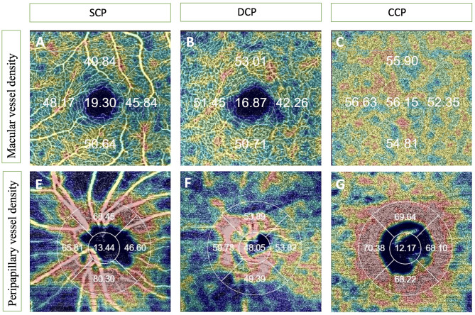
In this prospective study, a cohort of 31 healthy women with regular menstrual cycles was enrolled. SS-OCTA imaging was performed during three standardized menstrual phases: early follicular (day 3), ovulatory (day 14), and mid-luteal (day 21). Macular and peripapillary regions were evaluated using 3 × 3 mm and 4.5 × 4.5 mm scan protocols, respectively. Vessel density (VD) in the superficial capillary plexus (SCP), deep capillary plexus (DCP), and choriocapillaris (CC) was automatically quantified using the device's software. Choroidal thickness (CT) was manually measured in all quadrants, and the foveal avascular zone (FAZ) area was manually delineated by two independent graders. To control for diurnal variation, all measurements were conducted between 11:00 a.m. and 12:00 p.m.
A statistically significant decrease in macular CT was observed across the menstrual phases (p = 0.002), along with significant variations in peripapillary CT in the temporal and inferior quadrants (p = 0.004 for both). VD in the peripapillary region showed significant differences in the superior quadrant of the SCP (p = 0.040), the inferior quadrant of the DCP (p = 0.008), and in the temporal and inferior quadrants of the CC (p = 0.011 and p = 0.007, respectively). In the macula, CC-VD in the inferior quadrant differed significantly (p = 0.030). FAZ area remained stable throughout the cycle. Post-hoc analysis revealed significant differences between the early follicular, ovulatory, and mid-luteal phases.
Hormonal fluctuations throughout the menstrual cycle appear to influence both retinal and choroidal microvasculature, particularly in the choriocapillaris and peripapillary regions. These findings underscore the importance of considering menstrual phase when interpreting OCTA measurements in women of reproductive age, to improve diagnostic accuracy and consistency in clinical and research settings.
使用扫频光学相干断层扫描血管造影(SS-OCTA)研究月经周期相关激素波动对视网膜和脉络膜微血管系统的影响。
在这项前瞻性研究中,纳入了31名月经周期规律的健康女性队列。在三个标准化月经阶段进行SS-OCTA成像:卵泡早期(第3天)、排卵期(第14天)和黄体中期(第21天)。分别使用3×3mm和4.5×4.5mm扫描方案评估黄斑区和视乳头周围区域。使用设备软件自动定量浅表毛细血管丛(SCP)、深部毛细血管丛(DCP)和脉络膜毛细血管(CC)中的血管密度(VD)。在所有象限手动测量脉络膜厚度(CT),并由两名独立分级人员手动划定黄斑无血管区(FAZ)面积。为控制昼夜变化,所有测量均在上午11:00至12:00之间进行。
在月经周期各阶段观察到黄斑CT有统计学显著下降(p = 0.002),视乳头周围颞下象限CT也有显著变化(两者p = 0.004)。视乳头周围区域的VD在SCP上象限(p = 0.040)、DCP下象限(p = 0.008)以及CC的颞下象限(分别为p = 0.011和p = 0.007)有显著差异。在黄斑区,下象限的CC-VD有显著差异(p = 0.030)。FAZ面积在整个周期保持稳定。事后分析显示卵泡早期、排卵期和黄体中期之间存在显著差异。
月经周期中的激素波动似乎会影响视网膜和脉络膜微血管系统,特别是在脉络膜毛细血管和视乳头周围区域。这些发现强调了在解释育龄女性的OCTA测量结果时考虑月经周期阶段的重要性,以提高临床和研究环境中的诊断准确性和一致性。