Ophthalmology Unit, "Fondazione Policlinico Universitario A. Gemelli, IRCCS,", Largo A. Gemelli, 8, 00168, Rome, Italy.
Catholic University "Sacro Cuore,", Rome, Italy.
Graefes Arch Clin Exp Ophthalmol. 2024 Jul;262(7):2057-2065. doi: 10.1007/s00417-023-06346-0. Epub 2024 Feb 26.
The aim of our study was to evaluate changes in the retinal and choriocapillaris circulations in patients with hypothalamic amenorrhea.
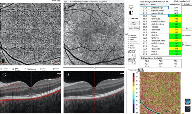
Prospective, cross-sectional observational study on 25 patients (50 eyes) diagnosed with hypothalamic amenorrhea and 25 age-matched healthy women. Optical coherence tomography angiography (OCTA) was used to evaluate the vessel density (VD) of superficial capillary plexus (SCP), deep capillary plexus (DCP), and choriocapillaris VD layers in whole 6.4 × 6.4-mm image and in fovea grid-based image. In patients' group, systemic parameters were collected: body mass index (BMI), endometrial rhyme thickness, follicle stimulating hormone (FSH), luteinizing hormone (LH), prolactin, insulin, and cortisol.
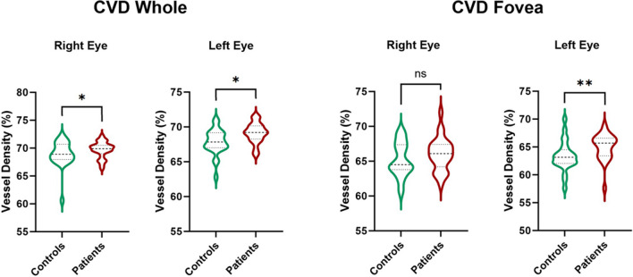
SCP and DCP did not show any statistical difference when comparing patients and controls (all p > 0.05). Differently, choriocapillaris VD in the whole region showed a non-significant tendency toward higher values in the patients group in both eyes (p = 0.038 for right eye [RE], p = 0.044 for left eye [LE]). Foveal choriocapillaris VD was higher in hypothalamic amenorrhea women vs. healthy controls (66.0 ± 2.4 vs. 63.7 ± 6.6%, p = 0.136 for RE; 65.0 ± 2.4 vs. 61.6 ± 7.0%, p = 0.005 for LE). Focusing on correlation with systemic parameters, SCP and DCP foveal density had a medium/high effect size with endometrial rhyme, along with DCP in the fovea area vs. cortisol and SCP in the whole area vs. FSH.
When comparing hypothalamic amenorrhea patients to healthy subjects, OCTA detected changes in the choriocapillaris layer, showing increased VD in the early stage of the systemic pathology, suggesting that microvascular "compaction" could be a first phase of hypoestrogenism adaptation.
本研究旨在评估下丘脑性闭经患者视网膜和脉络膜循环的变化。
对 25 例(50 只眼)确诊为下丘脑性闭经的患者和 25 名年龄匹配的健康女性进行前瞻性、横断面观察性研究。使用光学相干断层扫描血管造影(OCTA)评估浅层毛细血管丛(SCP)、深层毛细血管丛(DCP)和脉络膜毛细血管血管密度(VD)在整个 6.4×6.4mm 图像和黄斑网格图像中的分布。在患者组中,收集了系统参数:体重指数(BMI)、子宫内膜厚度、卵泡刺激素(FSH)、黄体生成素(LH)、催乳素、胰岛素和皮质醇。
SCP 和 DCP 在比较患者和对照组时没有统计学差异(所有 p>0.05)。不同的是,整个区域的脉络膜毛细血管 VD 在双眼患者组中呈现出非显著性增高趋势(右眼 p=0.038,左眼 p=0.044)。与健康对照组相比,下丘脑性闭经女性的黄斑区脉络膜毛细血管 VD 较高(右眼 66.0±2.4 对 63.7±6.6%,p=0.136;左眼 65.0±2.4 对 61.6±7.0%,p=0.005)。关于与系统参数的相关性,SCP 和 DCP 黄斑区密度与子宫内膜厚度呈中/高度相关,而 DCP 黄斑区密度与皮质醇呈正相关,SCP 全区域密度与 FSH 呈正相关。
与健康受试者相比,OCTA 在下丘脑性闭经患者中检测到脉络膜层的变化,表现为系统病理学早期的 VD 增加,提示微血管“紧缩”可能是低雌激素适应的第一阶段。