Shi Yingchao, Zhang Luming, Shu Xin, Zhang Keke, Yan Yuchao, Yuan Weizheng, Yu Yiting, Gong Yan
Ningbo Institute of Northwestern Polytechnical University, School of Mechanical Engineering, Northwestern Polytechnical University, Xi'an, Shaanxi 710072, China.
Key Laboratory of Micro/Nano Systems for Aerospace (Ministry of Education), Key Laboratory of Micro- and Nano-Electro-Mechanical Systems of Shaanxi Province, Northwestern Polytechnical University, Xi'an, Shaanxi 710072, China.
Biomed Opt Express. 2025 May 28;16(6):2482-2494. doi: 10.1364/BOE.563950. eCollection 2025 Jun 1.
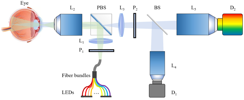
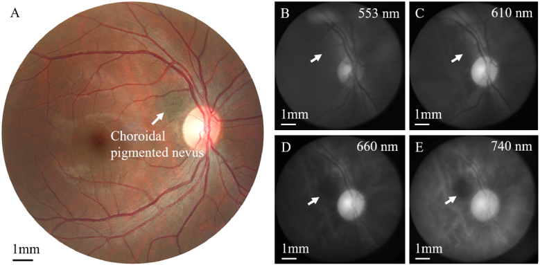
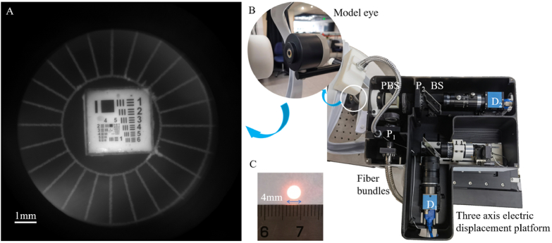
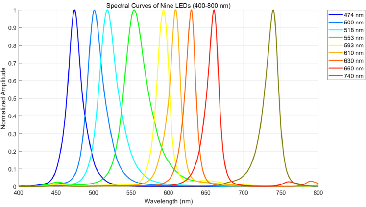
Fundus spectral imaging (FSI) integrates fundus photography with spectral techniques, providing both spatial and spectral information for retinal imaging. Whereas existing FSI systems have demonstrated advantages in structural and functional imaging, their widespread adoption is hindered by high costs and complex optical designs. To address these challenges, we propose a low-cost multispectral fundus camera with a simplified optical design, built from off-the-shelf optics, 3D-printed parts, and equipped with fiber-bundle-coupled multi-wavelength LED illumination source (470-740 nm). Additionally, the proposed multispectral imaging apparatus incorporates a coaxial non-separated polarization-based reflection suppression technique, using orthogonal polarizers to suppress corneal reflections without pupil-plane separation. To the best of our knowledge, this is the first application of such an architecture in the context of FSI. Experimental results demonstrate that the developed system achieves high-quality FSI under low-cost conditions, validating its feasibility as a practical solution. Clinical validation validates its diagnostic capability for diabetic retinopathy, choroidal pigmented nevus, and, notably, the first reported spectral imaging of peripapillary atrophy. The system achieves performance comparable to conventional color fundus photography while enabling superior diagnosis of deep fundus conditions such as choroidal lesions, offering a cost-effective and practical FSI solution for broader deployment in resource-limited settings.
眼底光谱成像(FSI)将眼底摄影与光谱技术相结合,为视网膜成像提供空间和光谱信息。尽管现有的FSI系统在结构和功能成像方面已展现出优势,但其高成本和复杂的光学设计阻碍了它们的广泛应用。为应对这些挑战,我们提出了一种低成本的多光谱眼底相机,其光学设计简化,由现成的光学器件、3D打印部件构建而成,并配备了光纤束耦合的多波长LED照明源(470 - 740纳米)。此外,所提出的多光谱成像设备采用了基于同轴非分离偏振的反射抑制技术,使用正交偏振器来抑制角膜反射,而无需在瞳孔平面进行分离。据我们所知,这是这种架构在FSI背景下的首次应用。实验结果表明,所开发的系统在低成本条件下实现了高质量的FSI,验证了其作为实际解决方案的可行性。临床验证证实了其对糖尿病视网膜病变、脉络膜色素痣的诊断能力,特别是首次报道的视乳头周围萎缩的光谱成像。该系统实现了与传统彩色眼底摄影相当的性能,同时能够对脉络膜病变等深部眼底疾病进行更优的诊断,为在资源有限的环境中更广泛地部署提供了一种经济高效且实用的FSI解决方案。