Department of Neuroscience-Ophthalmology, University of Padova, Padova, Italy.
Oftalmico Hospital, ASST Fatebenefratelli Sacco, Milano, Italy.
PLoS One. 2022 Aug 12;17(8):e0272318. doi: 10.1371/journal.pone.0272318. eCollection 2022.
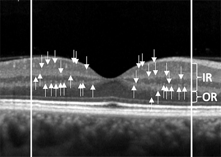
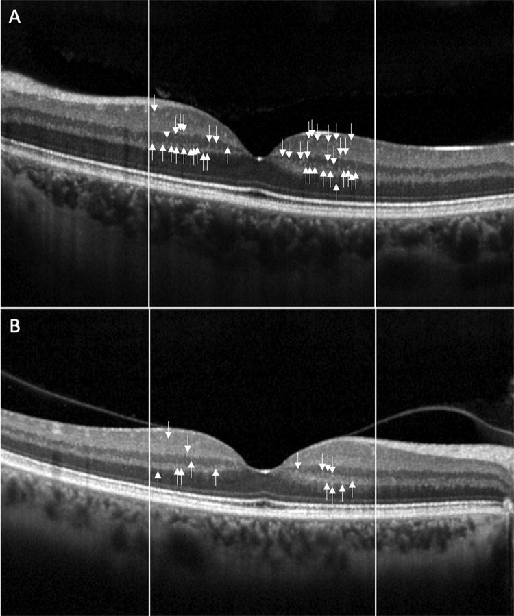
von Hippel-Lindau (VHL) disease is caused by a mutation of the VHL gene and characterized by the development of retinal hemangioblastomas (RH). Current pathophysiologic mechanisms of RH development and progression are still insufficient to predict RH behavior. VHL gene is involved in the cellular response to hypoxia and in many intracellular signaling pathways expressed both in angiogenesis and inflammation. Optical coherence tomography (OCT) allows to identify hyper-reflective retinal foci (HRF) known as aggregates of activated microglial cells as possible in vivo biomarker of local inflammation. The aim of the present study was to investigate the presence of HRF in patients with genetically confirmed VHL disease.
In this cross-sectional study, patients with VHL underwent complete ophthalmological examination and OCT with HRA + OCT Spectralis. HRF were manually identified and calculated in inner (IR), outer (OR) and full retina. Age-matched healthy subjects were enrolled as controls.
113 eyes of 63 VHL patients and 56 eyes of 28 healthy subjects were evaluated. HRF number was significantly higher in VHL than in controls in IR (28.06 ± 7.50 vs 25.25 ± 6.64, p = 0.042). No difference was observed in OR and in full retina (OR: 7.73 ± 2.59 vs 7.95 ± 2.51, p = 0.599; full retina: 35.79 ± 8.77 vs 33.20 ± 7.47, p = 0.093).
The increase of HRF, which mirror retinal microglial activation, characterizes VHL eyes. The role of activated microglia in the retina of VHL eyes needs to be better investigated, mainly considering local VHL disease manifestations.
von Hippel-Lindau(VHL)病是由 VHL 基因突变引起的,其特征是视网膜血管母细胞瘤(RH)的发展。目前,RH 发展和进展的病理生理机制仍不足以预测 RH 的行为。VHL 基因参与细胞对缺氧的反应,以及在血管生成和炎症中表达的许多细胞内信号通路。光学相干断层扫描(OCT)可识别高反射视网膜焦点(HRF),即激活的小胶质细胞聚集,作为局部炎症的潜在活体生物标志物。本研究旨在探讨遗传性 VHL 病患者中 HRF 的存在情况。
在这项横断面研究中,VHL 患者接受了全面的眼科检查和 HRA + OCT Spectralis 的 OCT 检查。手动识别和计算 HRF 在视网膜内(IR)、视网膜外(OR)和整个视网膜中的数量。招募年龄匹配的健康受试者作为对照组。
共评估了 63 例 VHL 患者的 113 只眼和 28 例健康对照者的 56 只眼。VHL 患者的 HRF 数量明显高于对照组的 IR(28.06±7.50 与 25.25±6.64,p=0.042)。OR 和全视网膜中未见差异(OR:7.73±2.59 与 7.95±2.51,p=0.599;全视网膜:35.79±8.77 与 33.20±7.47,p=0.093)。
HRF 的增加反映了视网膜小胶质细胞的激活,这是 VHL 眼的特征。需要进一步研究激活的小胶质细胞在 VHL 眼中的作用,主要考虑到局部 VHL 疾病的表现。