Kyawsoewin Maythwe, Manokawinchoke Jeeranan, Termkwanchareon Chutimon, Lwin Hnin Yu, Yaklai Sanicha, Limjeerajarus Nuttapol, Limjeerajarus Chalida Nakalekha, Egusa Hiroshi, Osathanon Thanaphum, Limraksasin Phoonsuk
Center of Excellence for Dental Stem Cell Biology, Faculty of Dentistry, Chulalongkorn University, Bangkok, Thailand.
Department of Anatomy, Faculty of Dentistry, Chulalongkorn University, Bangkok, Thailand.
Sci Rep. 2025 Jul 22;15(1):26589. doi: 10.1038/s41598-025-12323-w.
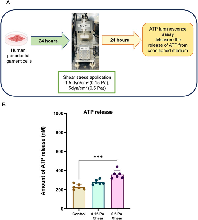
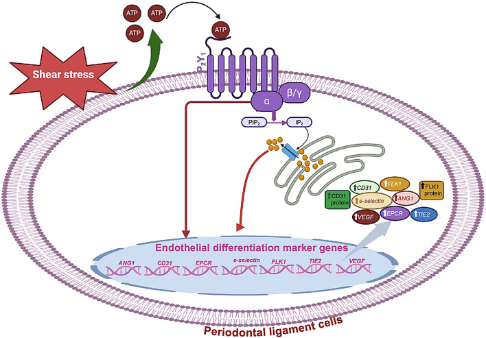
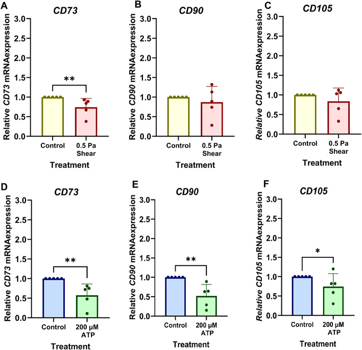
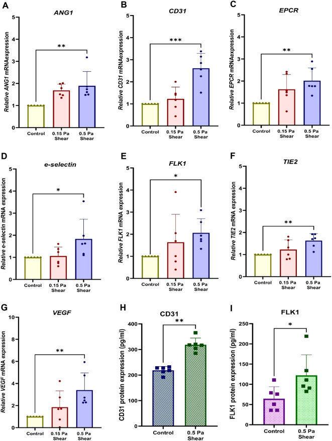
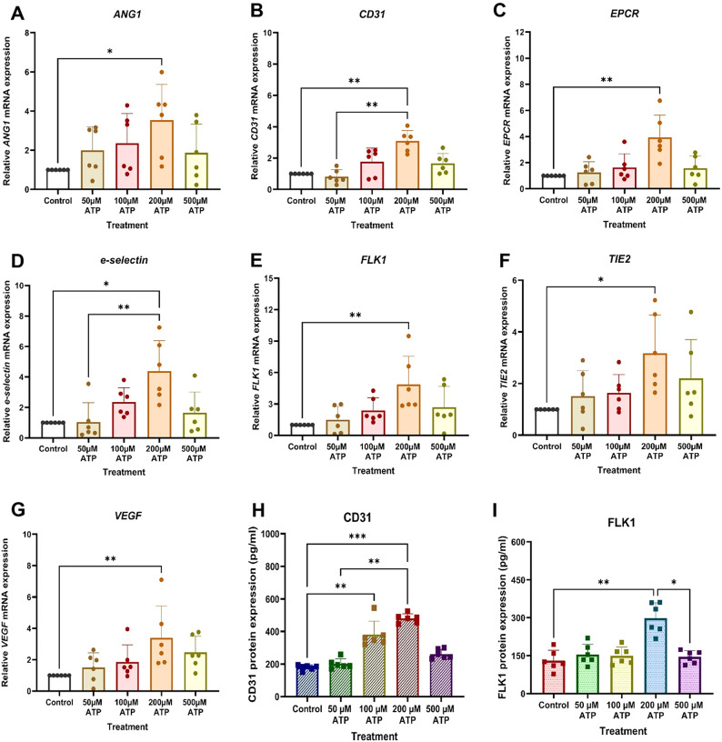
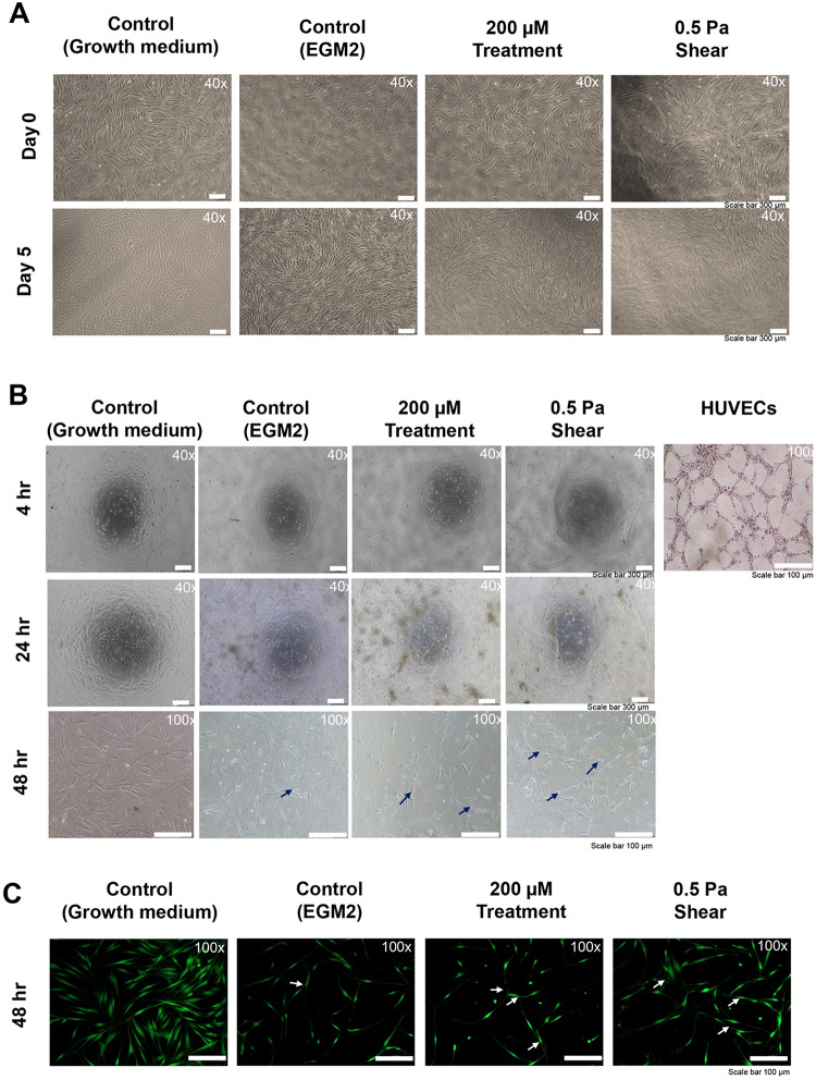
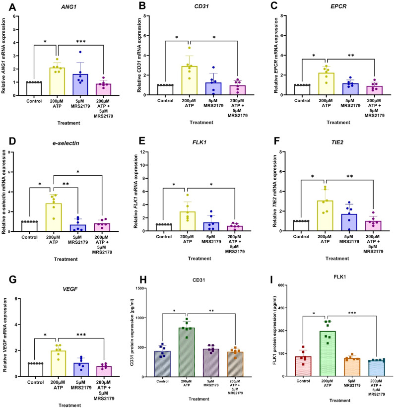
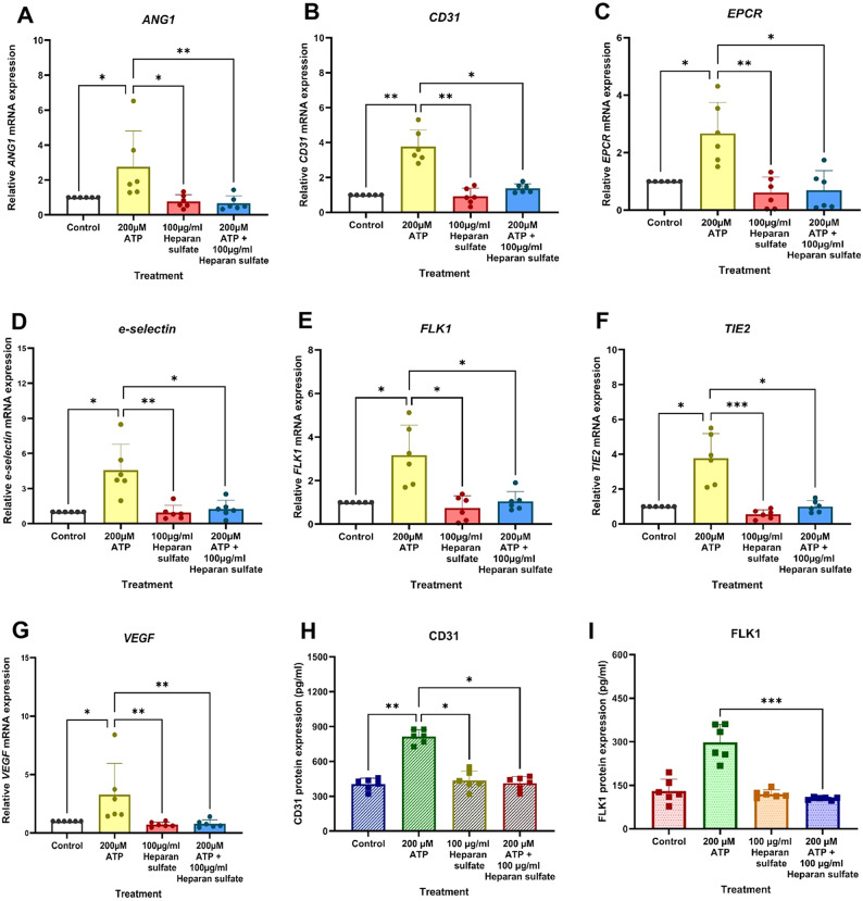
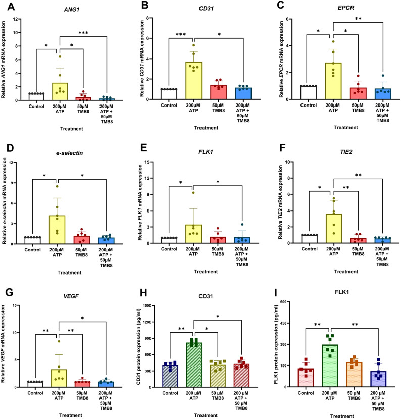
Mechanical forces stimulate human periodontal ligament stem cells (HPDLSCs) to release extracellular adenosine triphosphate (eATP). The eATP impacts various functions of HPDLSCs, i.e., immunosuppression and inflammation. eATP has been reported to promote the angiogenesis of pulmonary vascular endothelial cells. Shear force, one of the mechanical forces involved in orthodontic tooth movement, influences osteogenic differentiation and ECM remodeling of HPDLSCs. However, the relationship between shear force and the impact of eATP on endothelial differentiation and angiogenic characteristics of HPDLSCs remains unclear. This study aimed to determine the response of HPDLSCs on endothelial differentiation and angiogenic properties after shear stress loading and eATP treatment as well as explored the mechanism of eATP-involved in angiogenic responses of HPDLSCs. Shear stress application at 5 dyn/cm for 24 h stimulated the release of ATP by HPDLSCs. Both shear stress and 200 µM eATP promoted the expression of endothelial differentiation and angiogenic markers (ANG1, CD31, EPCR, e-selectin, FLK1, TIE2, VEGF). Blockade of specific PY receptor and intracellular calcium signaling attenuated eATP-induced expression of endothelial differentiation and angiogenic expression. Both shear stress and eATP have stimulatory effects on endothelial differentiation and angiogenesis in HPDLSCs through PY-intracellular calcium signaling.
机械力刺激人牙周膜干细胞(HPDLSCs)释放细胞外三磷酸腺苷(eATP)。eATP影响HPDLSCs的多种功能,即免疫抑制和炎症。据报道,eATP可促进肺血管内皮细胞的血管生成。剪切力是正畸牙齿移动过程中涉及的机械力之一,它影响HPDLSCs的成骨分化和细胞外基质重塑。然而,剪切力与eATP对HPDLSCs内皮分化和血管生成特性的影响之间的关系仍不清楚。本研究旨在确定剪切应力加载和eATP处理后HPDLSCs对内皮分化和血管生成特性的反应,并探讨eATP参与HPDLSCs血管生成反应的机制。以5达因/平方厘米的剪切应力施加24小时可刺激HPDLSCs释放ATP。剪切应力和200μM eATP均促进内皮分化和血管生成标志物(ANG1、CD31、EPCR、e-选择素、FLK1、TIE2、VEGF)的表达。特异性PY受体和细胞内钙信号的阻断减弱了eATP诱导的内皮分化和血管生成表达。剪切应力和eATP均通过PY-细胞内钙信号对HPDLSCs的内皮分化和血管生成具有刺激作用。