• 文献检索
  • 文档翻译
  • 深度研究
  • 学术资讯
  • Suppr Zotero 插件Zotero 插件
  • 邀请有礼
  • 套餐&价格
  • 历史记录
应用&插件
Suppr Zotero 插件Zotero 插件浏览器插件Mac 客户端Windows 客户端微信小程序
定价
高级版会员购买积分包购买API积分包
服务
文献检索文档翻译深度研究API 文档MCP 服务
关于我们
关于 Suppr公司介绍联系我们用户协议隐私条款
关注我们

Suppr 超能文献

核心技术专利:CN118964589B侵权必究
粤ICP备2023148730 号-1Suppr @ 2026

文献检索

告别复杂PubMed语法,用中文像聊天一样搜索,搜遍4000万医学文献。AI智能推荐,让科研检索更轻松。

立即免费搜索

文件翻译

保留排版,准确专业,支持PDF/Word/PPT等文件格式,支持 12+语言互译。

免费翻译文档

深度研究

AI帮你快速写综述,25分钟生成高质量综述,智能提取关键信息,辅助科研写作。

立即免费体验

关于扩散模型在微观图像和类微观图像分析中的应用的最新综述。

A state-of-the-art review of diffusion model applications for microscopic image and micro-alike image analysis.

作者信息

Liu Yan, Jiang Tao, Li Rui, Yuan Lingling, Grzegorzek Marcin, Li Chen, Li Xiaoyan

机构信息

College of Medicine and Biological Informaton Engineering, Northeastern University, Shenyang, China.

College of Intelligent Medicine, Chengdu University of Traditional Chinese Medicine, Chengdu, China.

出版信息

Front Med (Lausanne). 2025 Jul 16;12:1551894. doi: 10.3389/fmed.2025.1551894. eCollection 2025.

DOI:10.3389/fmed.2025.1551894
PMID:40740945
原文链接:https://pmc.ncbi.nlm.nih.gov/articles/PMC12309395/
Abstract

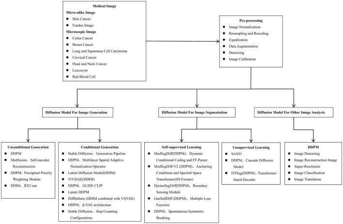
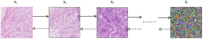
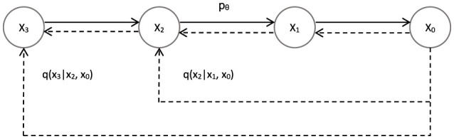
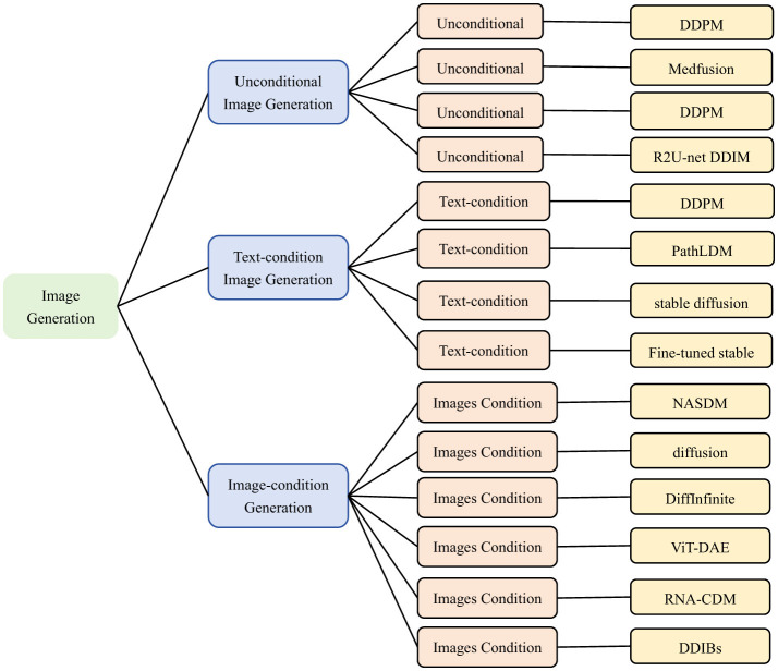
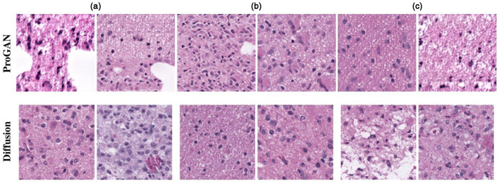
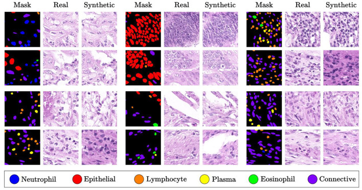
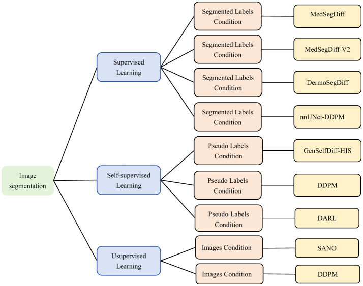
Diffusion models, a class of deep learning models based on probabilistic generative processes, progressively transform data into noise and then reconstruct the original data through an inverse process. Recently, diffusion models have gained attention in microscopic image analysis for their ability to process complex data, extract valuable information, and enhance image quality. This review provides an overview of diffusion models in microscopic images and micro-alike images, focusing on three commonly used models: DDPM, DDIM, and SDEs. We explore their applications in image generation, segmentation, denoising, classification, reconstruction and super-resolution. It shows their notable advantages, particularly in image generation and segmentation. Through simulating the imaging process of biological samples under the microscope, diffusion model can generate high-quality synthetic microscopic images. The generated images serve as a powerful tool for data augmentation when training deep learning models. Diffusion model also excels in microscopic image segmentation. It enables to accurately segment different cellular regions and tissue structures by simulating the interactions between pixels in an image. The review includes 31 papers, with 13 on image generation, nine on segmentation, and the remainder on other applications. We also discuss the strengths, limitations, and future directions for diffusion models in biomedical image processing.

摘要

扩散模型是一类基于概率生成过程的深度学习模型,它逐步将数据转换为噪声,然后通过逆过程重建原始数据。近年来,扩散模型因其处理复杂数据、提取有价值信息以及提高图像质量的能力而在微观图像分析中受到关注。本文综述了扩散模型在微观图像和类微观图像中的应用,重点介绍了三种常用模型:DDPM、DDIM和SDEs。我们探讨了它们在图像生成、分割、去噪、分类、重建和超分辨率方面的应用。结果表明,它们具有显著优势,尤其是在图像生成和分割方面。通过模拟生物样本在显微镜下的成像过程,扩散模型可以生成高质量的合成微观图像。生成的图像在训练深度学习模型时可作为强大的数据增强工具。扩散模型在微观图像分割方面也表现出色。它能够通过模拟图像中像素之间的相互作用,准确分割不同的细胞区域和组织结构。该综述包含31篇论文,其中13篇关于图像生成,9篇关于分割,其余涉及其他应用。我们还讨论了扩散模型在生物医学图像处理中的优势、局限性和未来发展方向。

https://cdn.ncbi.nlm.nih.gov/pmc/blobs/6693/12309395/4c31a1ced251/fmed-12-1551894-g0016.jpg
https://cdn.ncbi.nlm.nih.gov/pmc/blobs/6693/12309395/a4c0f9040149/fmed-12-1551894-g0001.jpg
https://cdn.ncbi.nlm.nih.gov/pmc/blobs/6693/12309395/e4e3ebb49943/fmed-12-1551894-g0002.jpg
https://cdn.ncbi.nlm.nih.gov/pmc/blobs/6693/12309395/c8ccb6380313/fmed-12-1551894-g0003.jpg
https://cdn.ncbi.nlm.nih.gov/pmc/blobs/6693/12309395/9f55c736436b/fmed-12-1551894-g0004.jpg
https://cdn.ncbi.nlm.nih.gov/pmc/blobs/6693/12309395/76e90ef3efd7/fmed-12-1551894-g0005.jpg
https://cdn.ncbi.nlm.nih.gov/pmc/blobs/6693/12309395/cc2b6c7eb2f0/fmed-12-1551894-g0006.jpg
https://cdn.ncbi.nlm.nih.gov/pmc/blobs/6693/12309395/d8033e37f4a0/fmed-12-1551894-g0007.jpg
https://cdn.ncbi.nlm.nih.gov/pmc/blobs/6693/12309395/45ac36452486/fmed-12-1551894-g0008.jpg
https://cdn.ncbi.nlm.nih.gov/pmc/blobs/6693/12309395/0f0b3951992b/fmed-12-1551894-g0009.jpg
https://cdn.ncbi.nlm.nih.gov/pmc/blobs/6693/12309395/4361ee24dc94/fmed-12-1551894-g0010.jpg
https://cdn.ncbi.nlm.nih.gov/pmc/blobs/6693/12309395/a942eb99ecab/fmed-12-1551894-g0011.jpg
https://cdn.ncbi.nlm.nih.gov/pmc/blobs/6693/12309395/47c9ca349150/fmed-12-1551894-g0012.jpg
https://cdn.ncbi.nlm.nih.gov/pmc/blobs/6693/12309395/06a7533ea7c6/fmed-12-1551894-g0013.jpg
https://cdn.ncbi.nlm.nih.gov/pmc/blobs/6693/12309395/192151ee46f6/fmed-12-1551894-g0014.jpg
https://cdn.ncbi.nlm.nih.gov/pmc/blobs/6693/12309395/5fbb8955a530/fmed-12-1551894-g0015.jpg
https://cdn.ncbi.nlm.nih.gov/pmc/blobs/6693/12309395/4c31a1ced251/fmed-12-1551894-g0016.jpg

相似文献

1
A state-of-the-art review of diffusion model applications for microscopic image and micro-alike image analysis.关于扩散模型在微观图像和类微观图像分析中的应用的最新综述。
Front Med (Lausanne). 2025 Jul 16;12:1551894. doi: 10.3389/fmed.2025.1551894. eCollection 2025.
2
Short-Term Memory Impairment短期记忆障碍
3
A medical image classification method based on self-regularized adversarial learning.基于自正则化对抗学习的医学图像分类方法。
Med Phys. 2024 Nov;51(11):8232-8246. doi: 10.1002/mp.17320. Epub 2024 Jul 30.
4
Influence of early through late fusion on pancreas segmentation from imperfectly registered multimodal magnetic resonance imaging.早期至晚期融合对来自配准不完善的多模态磁共振成像的胰腺分割的影响。
J Med Imaging (Bellingham). 2025 Mar;12(2):024008. doi: 10.1117/1.JMI.12.2.024008. Epub 2025 Apr 26.
5
Improving reconstruction of patient-specific abnormalities in AI-driven fast MRI with an individually adapted diffusion model.利用个体适配的扩散模型改进人工智能驱动的快速磁共振成像中患者特异性异常的重建。
Med Phys. 2025 Jul;52(7):e17955. doi: 10.1002/mp.17955.
6
Sexual Harassment and Prevention Training性骚扰与预防培训
7
Exploring the Potential of Electroencephalography Signal-Based Image Generation Using Diffusion Models: Integrative Framework Combining Mixed Methods and Multimodal Analysis.利用扩散模型探索基于脑电图信号的图像生成潜力:结合混合方法和多模态分析的综合框架
JMIR Med Inform. 2025 Jun 25;13:e72027. doi: 10.2196/72027.
8
Point-cloud segmentation with in-silico data augmentation for prostate cancer treatment.用于前列腺癌治疗的基于计算机模拟数据增强的点云分割
Med Phys. 2025 Apr 3. doi: 10.1002/mp.17815.
9
Full-dose whole-body PET synthesis from low-dose PET using high-efficiency denoising diffusion probabilistic model: PET consistency model.基于高效去噪扩散概率模型的低剂量全身 PET 全剂量合成:PET 一致性模型。
Med Phys. 2024 Aug;51(8):5468-5478. doi: 10.1002/mp.17068. Epub 2024 Apr 8.
10
Image quality evaluation in deep-learning-based CT noise reduction using virtual imaging trial methods: Contrast-dependent spatial resolution.基于深度学习的 CT 降噪中使用虚拟成像试验方法的图像质量评估:对比依赖性空间分辨率。
Med Phys. 2024 Aug;51(8):5399-5413. doi: 10.1002/mp.17029. Epub 2024 Mar 31.

本文引用的文献

1
Few-Shot Class-Incremental Learning for Classification and Object Detection: A Survey.用于分类和目标检测的少样本类别增量学习:综述
IEEE Trans Pattern Anal Mach Intell. 2025 Apr;47(4):2924-2945. doi: 10.1109/TPAMI.2025.3529038. Epub 2025 Mar 6.
2
Spontaneous breaking of symmetry in overlapping cell instance segmentation using diffusion models.使用扩散模型在重叠细胞实例分割中实现对称性的自发破缺
Biol Methods Protoc. 2024 Nov 9;9(1):bpae084. doi: 10.1093/biomethods/bpae084. eCollection 2024.
3
This Microtubule Does Not Exist: Super-Resolution Microscopy Image Generation by a Diffusion Model.
此微管不存在:基于扩散模型的超分辨率显微镜图像生成
Small Methods. 2025 Mar;9(3):e2400672. doi: 10.1002/smtd.202400672. Epub 2024 Oct 14.
4
GenSelfDiff-HIS: Generative Self-Supervision Using Diffusion for Histopathological Image Segmentation.GenSelfDiff-HIS:利用扩散进行组织病理学图像分割的生成式自监督方法
IEEE Trans Med Imaging. 2025 Feb;44(2):618-631. doi: 10.1109/TMI.2024.3453492. Epub 2025 Feb 4.
5
Diffusion-based deep learning method for augmenting ultrastructural imaging and volume electron microscopy.基于扩散的深度学习方法用于增强超微结构成像和体积电子显微镜技术。
Nat Commun. 2024 Jun 1;15(1):4677. doi: 10.1038/s41467-024-49125-z.
6
PathLDM: Text conditioned Latent Diffusion Model for Histopathology.PathLDM:用于组织病理学的文本条件潜扩散模型
IEEE Winter Conf Appl Comput Vis. 2024 Jan;2024:5170-5179. doi: 10.1109/wacv57701.2024.00510. Epub 2024 Apr 9.
7
Diffusion models in bioinformatics and computational biology.生物信息学和计算生物学中的扩散模型。
Nat Rev Bioeng. 2024 Feb;2(2):136-154. doi: 10.1038/s44222-023-00114-9. Epub 2023 Oct 27.
8
A visual-language foundation model for pathology image analysis using medical Twitter.一种使用医学推特进行病理学图像分析的视觉语言基础模型。
Nat Med. 2023 Sep;29(9):2307-2316. doi: 10.1038/s41591-023-02504-3. Epub 2023 Aug 17.
9
Reversing skin cancer adversarial examples by multiscale diffusive and denoising aggregation mechanism.通过多尺度扩散和去噪聚合机制来反转皮肤癌对抗样本。
Comput Biol Med. 2023 Sep;164:107310. doi: 10.1016/j.compbiomed.2023.107310. Epub 2023 Jul 31.
10
A multimodal comparison of latent denoising diffusion probabilistic models and generative adversarial networks for medical image synthesis.基于潜在去噪扩散概率模型和生成对抗网络的医学图像合成的多模态比较。
Sci Rep. 2023 Jul 26;13(1):12098. doi: 10.1038/s41598-023-39278-0.