• 文献检索
  • 文档翻译
  • 深度研究
  • 学术资讯
  • Suppr Zotero 插件Zotero 插件
  • 邀请有礼
  • 套餐&价格
  • 历史记录
应用&插件
Suppr Zotero 插件Zotero 插件浏览器插件Mac 客户端Windows 客户端微信小程序
定价
高级版会员购买积分包购买API积分包
服务
文献检索文档翻译深度研究API 文档MCP 服务
关于我们
关于 Suppr公司介绍联系我们用户协议隐私条款
关注我们

Suppr 超能文献

核心技术专利:CN118964589B侵权必究
粤ICP备2023148730 号-1Suppr @ 2026

文献检索

告别复杂PubMed语法,用中文像聊天一样搜索,搜遍4000万医学文献。AI智能推荐,让科研检索更轻松。

立即免费搜索

文件翻译

保留排版,准确专业,支持PDF/Word/PPT等文件格式,支持 12+语言互译。

免费翻译文档

深度研究

AI帮你快速写综述,25分钟生成高质量综述,智能提取关键信息,辅助科研写作。

立即免费体验

用于肺影像报告和数据系统(Lung-RADS)≥4A类结节良恶性鉴别的多模态列线图:氧增强零回波时间磁共振成像、CT影像组学和临床因素的整合

A multimodal nomogram for benign-malignant discrimination of lung-RADS ≥4A nodules: integration of oxygen enhanced zero echo time MRI, CT radiomics, and clinical factors.

作者信息

Yan Tiancai, Liu Ling, Li Yuxin, Qin Chunhui, Guan Haonan, Zhang Tong

机构信息

Department of Radiology, The Fourth Hospital of Harbin Medical University, Harbin, China.

GE Healthcare, MR Research China, Beijing, China.

出版信息

Front Oncol. 2025 Jul 21;15:1563073. doi: 10.3389/fonc.2025.1563073. eCollection 2025.

DOI:10.3389/fonc.2025.1563073
PMID:40761252
原文链接:https://pmc.ncbi.nlm.nih.gov/articles/PMC12318770/
Abstract

BACKGROUND AND OBJECTIVE

Lung-RADS ≥4A nodules require urgent intervention. Low-dose CT (LDCT), the primary screening tool, involves cumulative radiation exposure-critical for patients with serial scans. Oxygen-enhanced zero-echo time MRI (OE-ZTE-MRI) shows potential for lung nodule evaluation. However, its additive value when combined with CT radiomics and clinical factors for Lung-RADS ≥4A nodules remains unproven. This study aimed to develop a preoperative prediction model integrating OE-ZTE-MRI/CT radiomics and clinical factors for benign-malignant discrimination of Lung-RADS ≥4A nodules and compare its performance against single-modality models.

METHODS

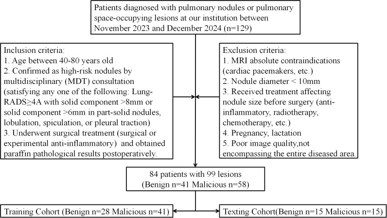
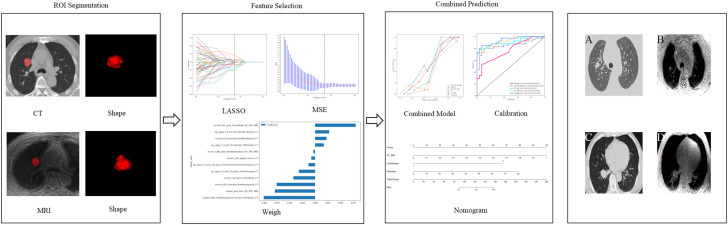
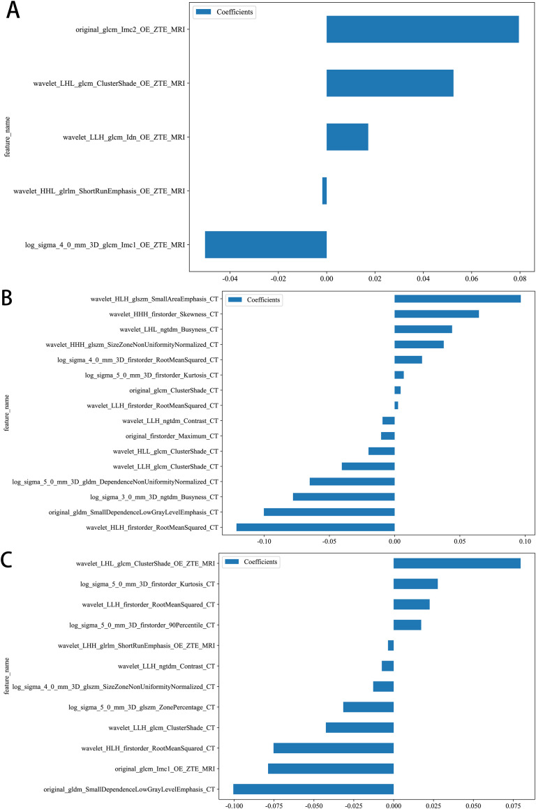
99 nodules from 84 prospectively enrolled patients undergoing both LDCT and OE-ZTE-MRI were included. Nodule boundaries were manually contoured as regions of interest (ROIs) on both modalities. Six machine learning classifiers were applied to radiomic features (extracted from LDCT and OE-ZTE-MRI) and clinical parameters (age, smoking history, nodule diameter, calcification, etc.). Model performance was evaluated using receiver operating characteristic (ROC) curves with area under the curve (AUC), complemented by decision curve analysis (DCA). Univariate and multivariate logistic regression identified independent predictors, which were incorporated into a final nomogram to visualize clinical-radiomic prediction.

RESULTS

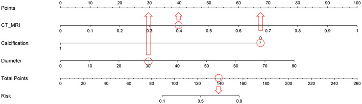
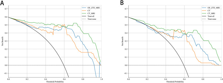
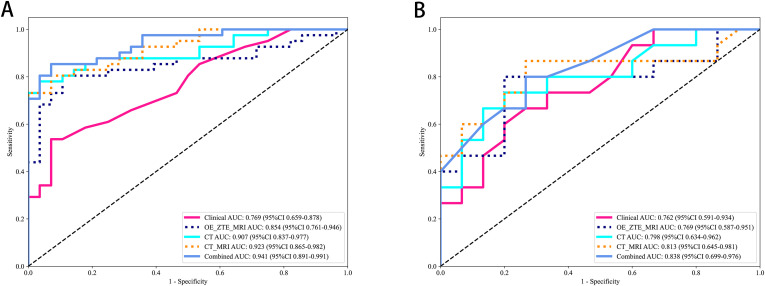
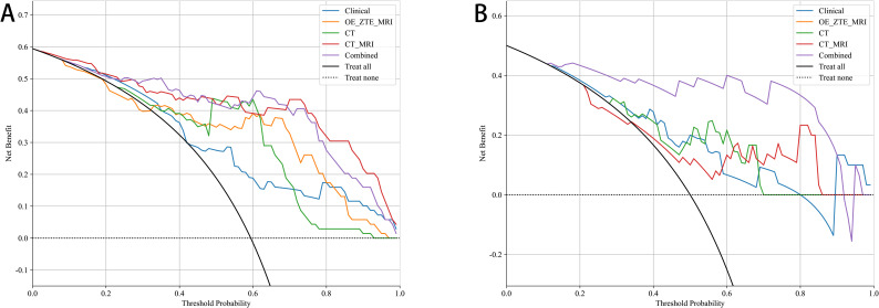
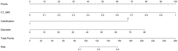
MRI model had a similar diagnostic performance to CT model (MRI . CT: training cohort AUC: 0.854 0.907; testing cohort AUC: 0.769 0.798). Multi-radiomics model achieved the highest diagnostic efficiency (train cohort AUC:0.923; testing cohort AUC: 0.813). Multivariate Logistic regression showed that nodule diameter (=0.005) and calcification (=0.029) were important factors affecting the benign and malignant nodules. The nomogram constructed by 3 models(CT/OE-ZTE-MRI/Clinical factors) achieved the best preoperative prediction performance for benign and malignant nodules (training cohort: AUC 0.941; testing cohort AUC:0.838).

CONCLUSION

The nomogram combining OE-ZTE-MRI/CT radiomics and clinical factors (nodule diameter, calcification) improves preoperative discrimination of Lung-RADS ≥4A nodules (AUC=0.838), outperforming single-modality models. This tool enables evidence-based triage, potentially reducing unnecessary invasive procedures.

摘要

背景与目的

Lung-RADS≥4A类结节需要紧急干预。低剂量CT(LDCT)作为主要的筛查工具,会使患者因多次扫描而累积接受辐射,这一点至关重要。氧增强零回波时间磁共振成像(OE-ZTE-MRI)显示出评估肺结节的潜力。然而,其与CT影像组学及临床因素相结合用于Lung-RADS≥4A类结节时的附加价值仍未得到证实。本研究旨在开发一种术前预测模型,该模型整合OE-ZTE-MRI/CT影像组学和临床因素,用于鉴别Lung-RADS≥4A类结节的良恶性,并将其性能与单模态模型进行比较。

方法

纳入了84例前瞻性招募的同时接受LDCT和OE-ZTE-MRI检查患者的99个结节。在两种检查方式下均手动勾勒出结节边界作为感兴趣区域(ROI)。将六种机器学习分类器应用于影像组学特征(从LDCT和OE-ZTE-MRI中提取)以及临床参数(年龄、吸烟史、结节直径、钙化等)。使用带有曲线下面积(AUC)的受试者工作特征(ROC)曲线评估模型性能,并辅以决策曲线分析(DCA)。单因素和多因素逻辑回归确定独立预测因素,并将其纳入最终的列线图以可视化临床影像组学预测。

结果

MRI模型的诊断性能与CT模型相似(MRI. CT:训练队列AUC:0.854 0.907;测试队列AUC:0.769 0.798)。多影像组学模型实现了最高的诊断效率(训练队列AUC:0.923;测试队列AUC:0.813)。多因素逻辑回归显示结节直径(=0.005)和钙化(=0.029)是影响结节良恶性的重要因素。由三种模型(CT/OE-ZTE-MRI/临床因素)构建的列线图对结节良恶性的术前预测性能最佳(训练队列:AUC 0.941;测试队列AUC:0.838)。

结论

结合OE-ZTE-MRI/CT影像组学和临床因素(结节直径、钙化)的列线图可改善对Lung-RADS≥4A类结节的术前鉴别(AUC = 0.838),优于单模态模型。该工具可实现基于证据的分诊,可能减少不必要的侵入性操作。

https://cdn.ncbi.nlm.nih.gov/pmc/blobs/d9e6/12318770/8141a740ca6a/fonc-15-1563073-g008.jpg
https://cdn.ncbi.nlm.nih.gov/pmc/blobs/d9e6/12318770/eacfdd62b962/fonc-15-1563073-g001.jpg
https://cdn.ncbi.nlm.nih.gov/pmc/blobs/d9e6/12318770/de716708f0a2/fonc-15-1563073-g002.jpg
https://cdn.ncbi.nlm.nih.gov/pmc/blobs/d9e6/12318770/f41c8b568122/fonc-15-1563073-g003.jpg
https://cdn.ncbi.nlm.nih.gov/pmc/blobs/d9e6/12318770/04de6a0dc31f/fonc-15-1563073-g004.jpg
https://cdn.ncbi.nlm.nih.gov/pmc/blobs/d9e6/12318770/c60492b15161/fonc-15-1563073-g005.jpg
https://cdn.ncbi.nlm.nih.gov/pmc/blobs/d9e6/12318770/ced34c8ac80e/fonc-15-1563073-g006.jpg
https://cdn.ncbi.nlm.nih.gov/pmc/blobs/d9e6/12318770/e2b26a6df947/fonc-15-1563073-g007.jpg
https://cdn.ncbi.nlm.nih.gov/pmc/blobs/d9e6/12318770/8141a740ca6a/fonc-15-1563073-g008.jpg

相似文献

1
A multimodal nomogram for benign-malignant discrimination of lung-RADS ≥4A nodules: integration of oxygen enhanced zero echo time MRI, CT radiomics, and clinical factors.用于肺影像报告和数据系统(Lung-RADS)≥4A类结节良恶性鉴别的多模态列线图:氧增强零回波时间磁共振成像、CT影像组学和临床因素的整合
Front Oncol. 2025 Jul 21;15:1563073. doi: 10.3389/fonc.2025.1563073. eCollection 2025.
2
Oxygen-Enhanced ZTE-MRI for Pulmonary Nodule Assessment: A Comparative Study with CT.用于肺结节评估的氧增强ZTE-MRI:与CT的对比研究
Acad Radiol. 2025 Aug;32(8):4903-4912. doi: 10.1016/j.acra.2025.04.036. Epub 2025 May 9.
3
Building radiomics models based on ACR TI-RADS combining clinical features for discriminating benign and malignant thyroid nodules.基于美国放射学会甲状腺影像报告和数据系统(ACR TI-RADS)构建联合临床特征的影像组学模型以鉴别甲状腺良恶性结节。
Front Endocrinol (Lausanne). 2025 Jul 21;16:1486920. doi: 10.3389/fendo.2025.1486920. eCollection 2025.
4
Radiomics Nomogram Based on Optimal Volume of Interest Derived from High-Resolution CT for Preoperative Prediction of IASLC Grading in Clinical IA Lung Adenocarcinomas: A Multi-Center, Large-Population Study.基于高分辨率 CT 最优感兴趣区体积的放射组学列线图预测临床 IA 期肺腺癌 IASLC 分级:多中心大样本研究。
Technol Cancer Res Treat. 2024 Jan-Dec;23:15330338241300734. doi: 10.1177/15330338241300734.
5
Radiomics features from whole thyroid gland tissue for prediction of cervical lymph node metastasis in the patients with papillary thyroid carcinoma.来自全甲状腺组织的影像组学特征用于预测甲状腺乳头状癌患者的颈部淋巴结转移。
J Cancer Res Clin Oncol. 2023 Nov;149(14):13005-13016. doi: 10.1007/s00432-023-05184-1. Epub 2023 Jul 19.
6
Nomogram based on radiomics and CT features for predicting visceral pleural invasion of invasive adenocarcinoma ≤ 2 cm: A multicenter study.基于影像组学和CT特征的列线图预测直径≤2 cm浸润性腺癌脏层胸膜侵犯:一项多中心研究
Eur J Radiol. 2025 Jun 12;190:112227. doi: 10.1016/j.ejrad.2025.112227.
7
Research on the application of distinguishing between benign and malignant breast nodules using MRI and US radiomics.利用MRI和超声影像组学鉴别乳腺良恶性结节的应用研究
Front Oncol. 2025 Jul 16;15:1630583. doi: 10.3389/fonc.2025.1630583. eCollection 2025.
8
Multimodal nomogram integrating deep learning radiomics and hemodynamic parameters for early prediction of post-craniotomy intracranial hypertension.整合深度学习影像组学和血流动力学参数的多模态列线图用于开颅术后颅内高压的早期预测
Sci Rep. 2025 Jul 2;15(1):23595. doi: 10.1038/s41598-025-09115-7.
9
Machine learning-based radiomics for differentiating lung cancer subtypes in brain metastases using CE-T1WI.基于机器学习的影像组学在使用对比增强T1加权成像鉴别脑转移瘤中肺癌亚型的应用
Front Oncol. 2025 Jun 19;15:1599882. doi: 10.3389/fonc.2025.1599882. eCollection 2025.
10
Peritumoral Radiomic Features on CT for Differential Diagnosis in Small-Cell Lung Cancer: Potential for Surgical Decision-Making.CT上小细胞肺癌鉴别诊断的瘤周放射组学特征:手术决策的潜力
Cancer Control. 2025 Jan-Dec;32:10732748251351754. doi: 10.1177/10732748251351754. Epub 2025 Jun 16.

本文引用的文献

1
[Chinese expert consensus on diagnosis and treatment of pulmonary nodules(2024)].《肺结节诊治中国专家共识(2024年版)》
Zhonghua Jie He He Hu Xi Za Zhi. 2024 Aug 12;47(8):716-729. doi: 10.3760/cma.j.cn112147-20231109-00300.
2
CT and MRI radiomic features of lung cancer (NSCLC): comparison and software consistency.肺癌(NSCLC)的 CT 和 MRI 影像组学特征:比较和软件一致性。
Eur Radiol Exp. 2024 Jun 17;8(1):71. doi: 10.1186/s41747-024-00468-8.
3
Assessment of Lung Nodule Detection and Lung CT Screening Reporting and Data System Classification Using Zero Echo Time Pulmonary MRI.
使用零回波时间肺部MRI评估肺结节检测及肺部CT筛查报告和数据系统分类
J Magn Reson Imaging. 2025 Feb;61(2):822-829. doi: 10.1002/jmri.29388. Epub 2024 Apr 11.
4
Global cancer statistics 2022: GLOBOCAN estimates of incidence and mortality worldwide for 36 cancers in 185 countries.2022 年全球癌症统计数据:全球 185 个国家和地区 36 种癌症的发病率和死亡率全球估计数。
CA Cancer J Clin. 2024 May-Jun;74(3):229-263. doi: 10.3322/caac.21834. Epub 2024 Apr 4.
5
Development of a combined radiomics and CT feature-based model for differentiating malignant from benign subcentimeter solid pulmonary nodules.开发一种联合放射组学和 CT 特征的模型,用于区分亚厘米实性肺结节的良恶性。
Eur Radiol Exp. 2024 Jan 17;8(1):8. doi: 10.1186/s41747-023-00400-6.
6
Clinical Manifestations and Management of Fibrotic Pulmonary Sarcoidosis.纤维化型结节病的临床表现与管理
J Clin Med. 2023 Dec 31;13(1):241. doi: 10.3390/jcm13010241.
7
Relationship between advanced lung cancer inflammation index and long-term all-cause, cardiovascular, and cancer mortality among type 2 diabetes mellitus patients: NHANES, 1999-2018.2 型糖尿病患者晚期肺癌炎症指数与全因、心血管和癌症死亡的关系:NHANES,1999-2018。
Front Endocrinol (Lausanne). 2023 Nov 28;14:1298345. doi: 10.3389/fendo.2023.1298345. eCollection 2023.
8
A Review of Advances in the Surgical Treatment of Coronary Heart Disease and Lung Cancer.冠状动脉心脏病与肺癌手术治疗进展述评。
Heart Surg Forum. 2023 Oct 30;26(5):E656-E665. doi: 10.59958/hsf.5877.
9
Feasibility of UTE-MRI-based radiomics model for prediction of histopathologic subtype of lung adenocarcinoma: in comparison with CT-based radiomics model.基于 UTE-MRI 的放射组学模型预测肺腺癌组织病理亚型的可行性:与 CT 基于放射组学模型的比较。
Eur Radiol. 2024 May;34(5):3422-3430. doi: 10.1007/s00330-023-10302-1. Epub 2023 Oct 16.
10
Baseline whole-lung CT features deriving from deep learning and radiomics: prediction of benign and malignant pulmonary ground-glass nodules.基于深度学习和影像组学的基线全肺CT特征:良性和恶性肺磨玻璃结节的预测
Front Oncol. 2023 Aug 17;13:1255007. doi: 10.3389/fonc.2023.1255007. eCollection 2023.