August Ari H, Wibbelsman Turner D, Massenzio Erik, Thornton Sarah E, Peričić Danijel J
Department of Pathology, Wills Eye Hospital, 840 Walnut Street, Philadelphia, PA, 19107, USA.
Department of Translational Ophthalmology, 840 Walnut Street, Philadelphia, PA, 19107, USA.
Am J Ophthalmol Case Rep. 2025 Jul 22;39:102395. doi: 10.1016/j.ajoc.2025.102395. eCollection 2025 Sep.
Orbital color Doppler imaging (CDI) is useful in the evaluation of sudden monocular vision loss, providing information on etiology which may guide management. We present two cases of amaurosis fugax progressing to retinal artery occlusion (RAO) associated with migration of a hyperechoic particle within the central retinal artery (CRA) and altered vascular dynamics found on CDI.
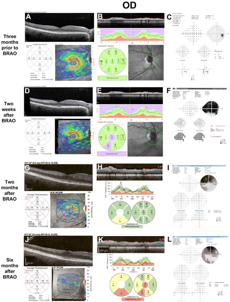
Both patients presented with amaurosis fugax, and CDI revealed a hyperechoic particle 2.8 mm from the optic nerve head in both patients. Patient 1 was found to have severe aortic stenosis and a thoracic aortic aneurysm and was managed with dual antiplatelet therapy (DAPT) while awaiting evaluation for cardiothoracic surgical repair. Ten days later, Patient 1 returned with a central RAO, and a repeat CDI showed a 1.0 mm anterior migration of the embolus with reduced CRA blood velocity and an increased resistivity index. Patient 2 was managed with DAPT and oral corticosteroids, but symptoms recurred during steroid tapering which necessitated a prolonged course of steroids. Systemic complications required reduction of steroid dosing, and the patient developed a branch RAO six months after initial presentation. Repeat CDI revealed a 0.9 mm anterior migration of the embolus, with increased CRA blood velocity and resistivity index. Systemic thrombolysis with tissue plasminogen activator and resumption of steroids did not result in visual improvement in Patient 2.
The presence of a hyperechoic particle in the CRA on CDI can be seen with amaurosis fugax, and anterior migration with subsequent alterations in CDI parameters may correlate with clinical progression to embolic retinal ischemia. Visualization of an embolus may predict nonresponse to thrombolytic or anticoagulation-based treatment.
眼眶彩色多普勒成像(CDI)有助于评估突然发生的单眼视力丧失,提供有关病因的信息,从而指导治疗。我们报告两例一过性黑矇进展为视网膜动脉阻塞(RAO)的病例,其与视网膜中央动脉(CRA)内高回声颗粒的移动以及CDI发现的血管动力学改变有关。
两名患者均表现为一过性黑矇,CDI显示两名患者在距视神经乳头2.8 mm处均有一个高回声颗粒。患者1被发现患有严重主动脉瓣狭窄和胸主动脉瘤,在等待心胸外科手术修复评估期间接受双重抗血小板治疗(DAPT)。十天后,患者1因中央型RAO复诊,重复CDI显示栓子向前移动1.0 mm,CRA血流速度降低,阻力指数增加。患者2接受DAPT和口服糖皮质激素治疗,但在激素减量过程中症状复发,这需要延长激素疗程。全身并发症需要减少激素剂量,患者在初次就诊六个月后发生分支型RAO。重复CDI显示栓子向前移动0.9 mm,CRA血流速度和阻力指数增加。患者2接受组织纤溶酶原激活剂全身溶栓并恢复使用激素后视力未改善。
CDI显示CRA内存在高回声颗粒可见于一过性黑矇,栓子向前移动及随后CDI参数的改变可能与栓塞性视网膜缺血的临床进展相关。栓子的可视化可能预示对溶栓或抗凝治疗无反应。