Wu Jiayi, Heller Stefan, Matern Maggie S
Department of Otolaryngology - Head and Neck Surgery, Stanford University School of Medicine, Stanford, CA 94304, USA; Institute for Stem Cell Biology & Regenerative Medicine, Stanford University School of Medicine, Stanford, CA 94304, USA.
Department of Otolaryngology - Head and Neck Surgery, Stanford University School of Medicine, Stanford, CA 94304, USA; Institute for Stem Cell Biology & Regenerative Medicine, Stanford University School of Medicine, Stanford, CA 94304, USA.
STAR Protoc. 2025 Aug 14;6(3):104032. doi: 10.1016/j.xpro.2025.104032.
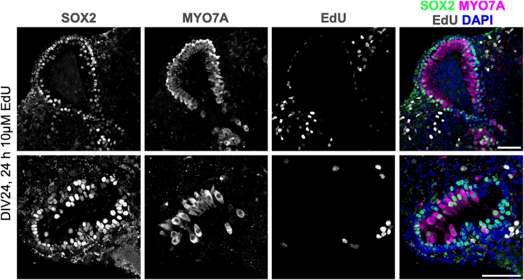
Inner ear organoids represent a potentially inexhaustible source of otic tissues, including sensory hair cells and supporting cells, for in vitro manipulation. Here, we present a protocol for labeling S-phase entry of cells in inner ear organoids using 5-ethynyl-2'-deoxyuridine (EdU), followed by fixation and vibratome sectioning. Nuclear EdU is then detected alongside protein markers of interest via immunofluorescence. This workflow enables the visualization of cell and tissue morphologies within developing organoids and assessment of how different manipulations affect cell proliferation. For complete details on the use and execution of this protocol, please refer to Matern et al..
内耳类器官是用于体外操作的耳组织(包括感觉毛细胞和支持细胞)潜在的无穷来源。在此,我们展示了一种使用5-乙炔基-2'-脱氧尿苷(EdU)标记内耳类器官中细胞进入S期的方案,随后进行固定和振动切片。然后通过免疫荧光检测细胞核中的EdU以及感兴趣的蛋白质标志物。该工作流程能够可视化发育中的类器官内的细胞和组织形态,并评估不同操作如何影响细胞增殖。有关此方案使用和执行的完整详细信息,请参考马特恩等人的文献。