• 文献检索
  • 文档翻译
  • 深度研究
  • 学术资讯
  • Suppr Zotero 插件Zotero 插件
  • 邀请有礼
  • 套餐&价格
  • 历史记录
应用&插件
Suppr Zotero 插件Zotero 插件浏览器插件Mac 客户端Windows 客户端微信小程序
定价
高级版会员购买积分包购买API积分包
服务
文献检索文档翻译深度研究API 文档MCP 服务
关于我们
关于 Suppr公司介绍联系我们用户协议隐私条款
关注我们

Suppr 超能文献

核心技术专利:CN118964589B侵权必究
粤ICP备2023148730 号-1Suppr @ 2026

文献检索

告别复杂PubMed语法,用中文像聊天一样搜索,搜遍4000万医学文献。AI智能推荐,让科研检索更轻松。

立即免费搜索

文件翻译

保留排版,准确专业,支持PDF/Word/PPT等文件格式,支持 12+语言互译。

免费翻译文档

深度研究

AI帮你快速写综述,25分钟生成高质量综述,智能提取关键信息,辅助科研写作。

立即免费体验

通过单细胞转录组学和多组学大数据分析揭示的宫颈癌中PTK6介导的免疫特征

PTK6 mediated immune signatures revealed by single cell transcriptomic and multi omics big data analysis in cervical cancer.

作者信息

Zhao Fen, Zhong Huanxin, You Lifang, Du Yi, Huang Changchang

机构信息

Department of Gynecology, First People's Hospital of Linping District, No. 369 Yingbin Road, Nanyuan Subdistrict, Linping District, Hangzhou, Zhejiang, China.

出版信息

Discov Oncol. 2025 Aug 16;16(1):1566. doi: 10.1007/s12672-025-03365-7.

DOI:10.1007/s12672-025-03365-7
PMID:40818002
原文链接:https://pmc.ncbi.nlm.nih.gov/articles/PMC12357816/
Abstract

BACKGROUND

Cervical cancer exhibits heterogeneous clinical outcomes, requiring improved prognostic tools. Single-cell RNA sequencing enables high-resolution analysis of tumor microenvironment cellular heterogeneity. This study developed a prognostic model for cervical cancer through single-cell transcriptomic analysis and immune infiltration characterization, focusing on PTK6 as a key biomarker.

METHODS

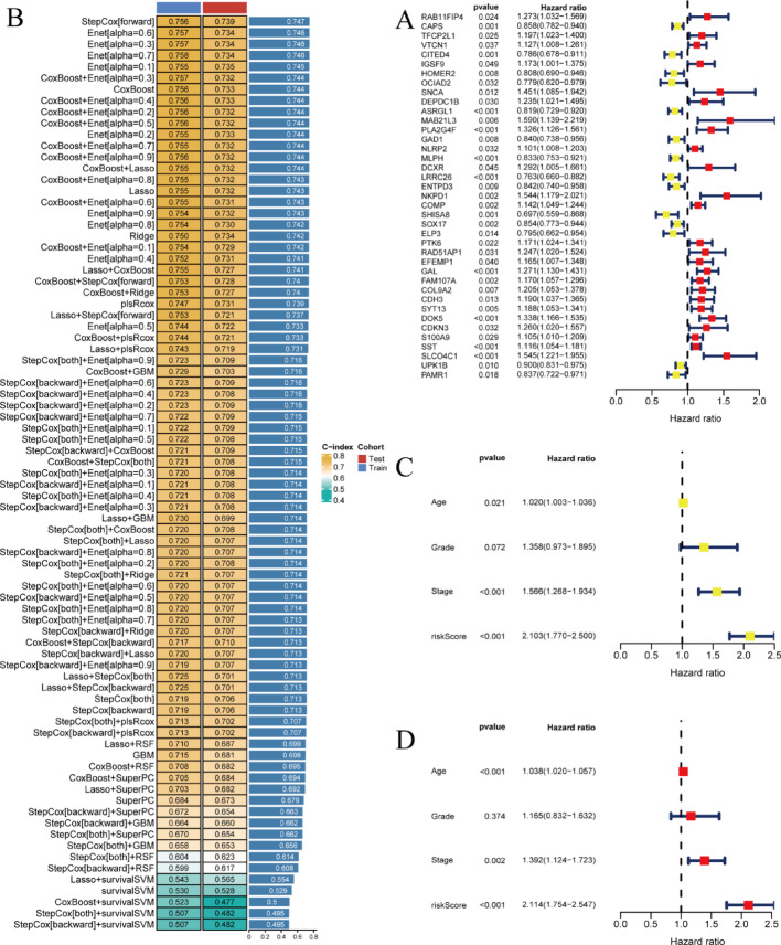
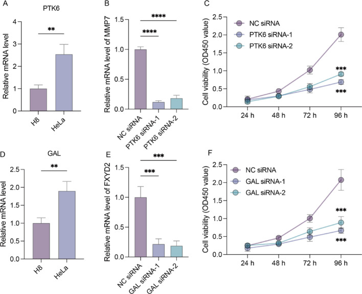
We analyzed TCGA and GEO transcriptomic data with single-cell RNA sequencing datasets. Fifteen machine learning algorithms constructed prognostic models using immune infiltration-related genes. Single-cell analysis employed Seurat for cell clustering and annotation. PTK6 expression was validated in H8 and HeLa cell lines via RT-qPCR and siRNA knockdown experiments.

RESULTS

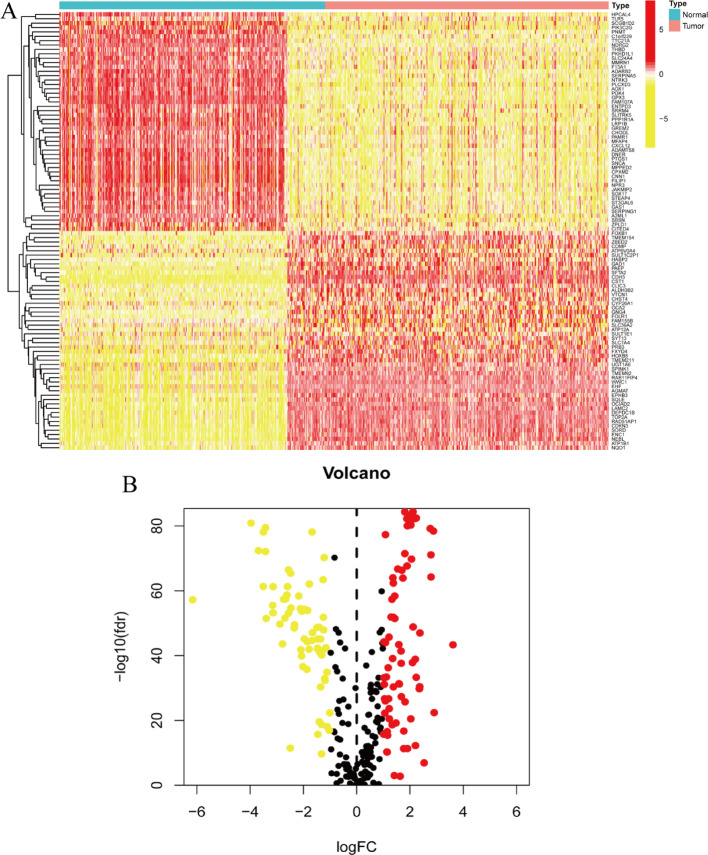
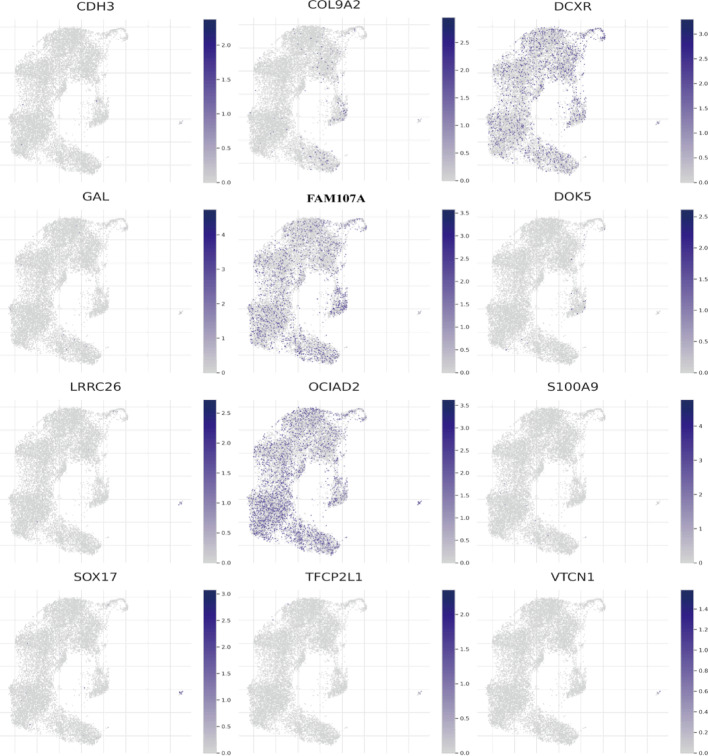
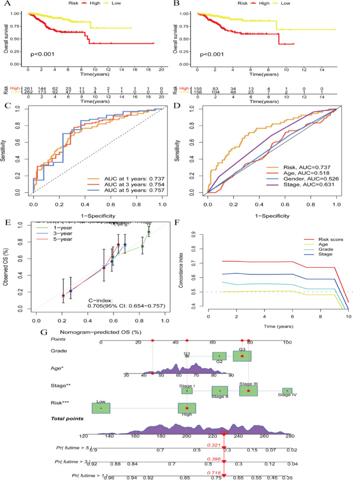
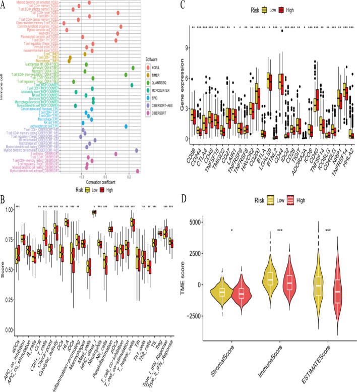
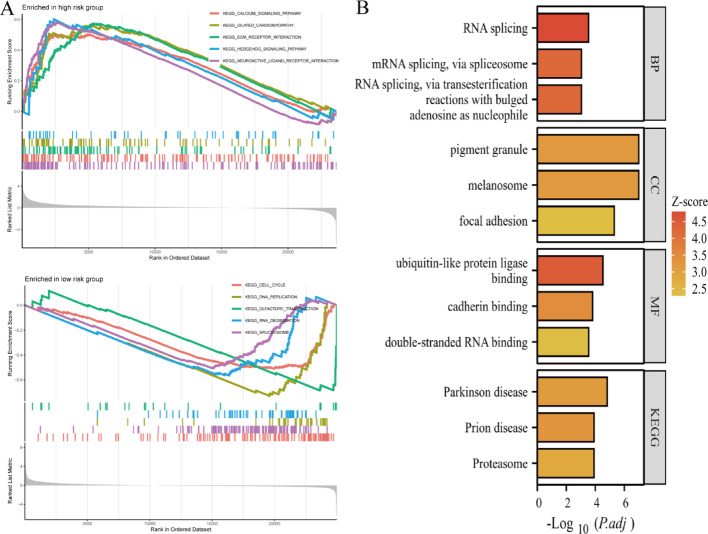
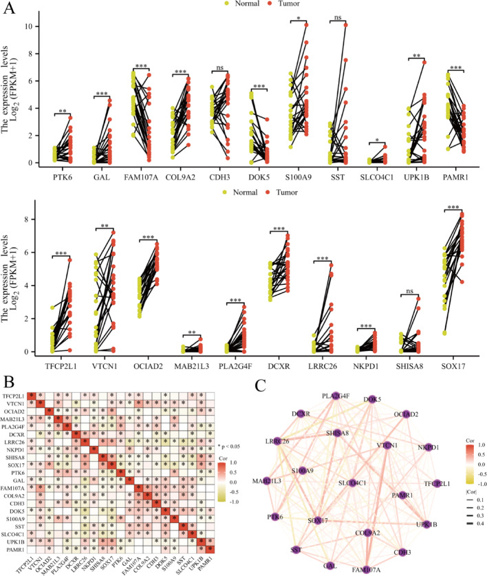
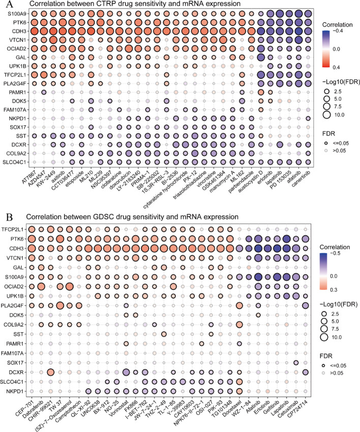
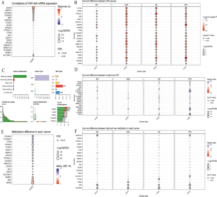
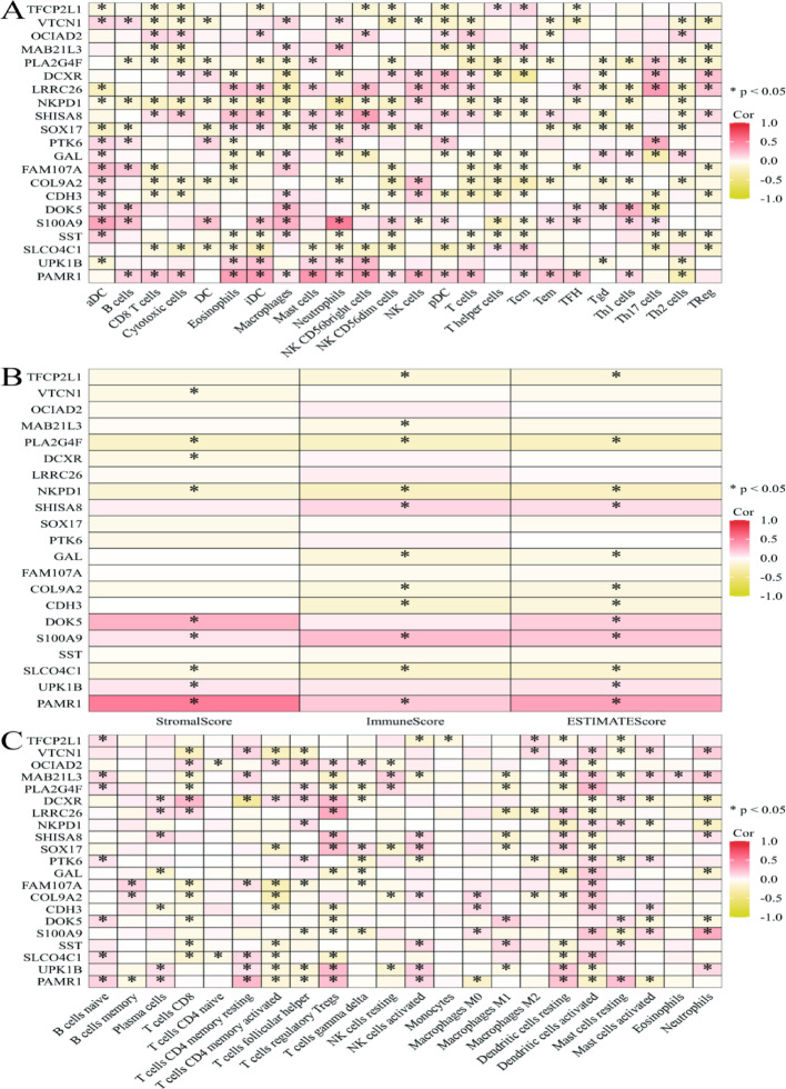
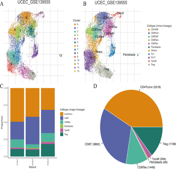
Single-cell sequencing revealed distinct cellular populations including CD8T cells, CD4Tconv cells, and fibroblasts. The prognostic model achieved excellent performance with AUC values of 0.737-0.757 across 1-5 years. PTK6 showed significantly elevated expression in tumors and strong correlations with immune infiltration. Single-cell analysis confirmed PTK6 expression across multiple cell types. Functional validation demonstrated that PTK6 knockdown reduced HeLa cell proliferation, confirming its oncogenic role.

CONCLUSION

PTK6 emerges as a critical immune infiltration-related prognostic biomarker through single-cell transcriptomic analysis.

摘要

背景

宫颈癌表现出异质性的临床结果,需要改进预后工具。单细胞RNA测序能够对肿瘤微环境细胞异质性进行高分辨率分析。本研究通过单细胞转录组分析和免疫浸润特征分析,开发了一种针对宫颈癌的预后模型,重点关注PTK6作为关键生物标志物。

方法

我们使用单细胞RNA测序数据集分析了TCGA和GEO转录组数据。15种机器学习算法使用免疫浸润相关基因构建预后模型。单细胞分析采用Seurat进行细胞聚类和注释。通过RT-qPCR和siRNA敲低实验在H8和HeLa细胞系中验证PTK6表达。

结果

单细胞测序揭示了不同的细胞群体,包括CD8T细胞、CD4Tconv细胞和成纤维细胞。该预后模型在1至5年期间的AUC值为0.737 - 0.757,表现出色。PTK6在肿瘤中表达显著升高,且与免疫浸润密切相关。单细胞分析证实了PTK6在多种细胞类型中的表达。功能验证表明,PTK6敲低可降低HeLa细胞增殖,证实了其致癌作用。

结论

通过单细胞转录组分析,PTK6成为一种关键的免疫浸润相关预后生物标志物。

https://cdn.ncbi.nlm.nih.gov/pmc/blobs/4059/12357816/a7817947d22c/12672_2025_3365_Fig12_HTML.jpg
https://cdn.ncbi.nlm.nih.gov/pmc/blobs/4059/12357816/dab392d636ad/12672_2025_3365_Fig1_HTML.jpg
https://cdn.ncbi.nlm.nih.gov/pmc/blobs/4059/12357816/adb02aae982f/12672_2025_3365_Fig2_HTML.jpg
https://cdn.ncbi.nlm.nih.gov/pmc/blobs/4059/12357816/d4a6f710fb4e/12672_2025_3365_Fig3_HTML.jpg
https://cdn.ncbi.nlm.nih.gov/pmc/blobs/4059/12357816/9aee0c6651c1/12672_2025_3365_Fig4_HTML.jpg
https://cdn.ncbi.nlm.nih.gov/pmc/blobs/4059/12357816/6a0c8aa8ba92/12672_2025_3365_Fig5_HTML.jpg
https://cdn.ncbi.nlm.nih.gov/pmc/blobs/4059/12357816/a17c1ea25719/12672_2025_3365_Fig6_HTML.jpg
https://cdn.ncbi.nlm.nih.gov/pmc/blobs/4059/12357816/f3304a7145a4/12672_2025_3365_Fig7_HTML.jpg
https://cdn.ncbi.nlm.nih.gov/pmc/blobs/4059/12357816/227d0ed6e33d/12672_2025_3365_Fig8_HTML.jpg
https://cdn.ncbi.nlm.nih.gov/pmc/blobs/4059/12357816/44e6fbfcf24b/12672_2025_3365_Fig9_HTML.jpg
https://cdn.ncbi.nlm.nih.gov/pmc/blobs/4059/12357816/8af0f86b1969/12672_2025_3365_Fig10_HTML.jpg
https://cdn.ncbi.nlm.nih.gov/pmc/blobs/4059/12357816/b3054adc8ca4/12672_2025_3365_Fig11_HTML.jpg
https://cdn.ncbi.nlm.nih.gov/pmc/blobs/4059/12357816/a7817947d22c/12672_2025_3365_Fig12_HTML.jpg

相似文献

1
PTK6 mediated immune signatures revealed by single cell transcriptomic and multi omics big data analysis in cervical cancer.通过单细胞转录组学和多组学大数据分析揭示的宫颈癌中PTK6介导的免疫特征
Discov Oncol. 2025 Aug 16;16(1):1566. doi: 10.1007/s12672-025-03365-7.
2
Bioinformatics-based prognostic value and functional validation of PTK6 in cutaneous melanoma.基于生物信息学的PTK6在皮肤黑色素瘤中的预后价值及功能验证
Front Oncol. 2025 Jul 30;15:1555302. doi: 10.3389/fonc.2025.1555302. eCollection 2025.
3
SLC3A2 as a key anoikis-related gene for prognosis and tumor microenvironment remodeling in melanoma.SLC3A2作为黑色素瘤预后和肿瘤微环境重塑的关键失巢凋亡相关基因。
Discov Oncol. 2025 Jul 11;16(1):1306. doi: 10.1007/s12672-025-03125-7.
4
Interplay between tumor mutation burden and the tumor microenvironment predicts the prognosis of pan-cancer anti-PD-1/PD-L1 therapy.肿瘤突变负荷与肿瘤微环境之间的相互作用可预测泛癌抗PD-1/PD-L1治疗的预后。
Front Immunol. 2025 Jul 24;16:1557461. doi: 10.3389/fimmu.2025.1557461. eCollection 2025.
5
Multi-gene anoikis signature combined with tumor microenvironment stratifies prognosis in cutaneous melanoma with experimental validation of PTK6.多基因失巢凋亡特征联合肿瘤微环境对皮肤黑色素瘤的预后进行分层,并对PTK6进行了实验验证。
Gene. 2025 Sep 20;966:149686. doi: 10.1016/j.gene.2025.149686. Epub 2025 Jul 26.
6
Identification of novel molecular subtypes and construction of a prognostic signature via multi-omics analysis and machine learning in lung adenocarcinoma.通过多组学分析和机器学习在肺腺癌中鉴定新的分子亚型并构建预后特征
Front Oncol. 2025 Jul 21;15:1590216. doi: 10.3389/fonc.2025.1590216. eCollection 2025.
7
Senescent fibroblasts secrete CTHRC1 to promote cancer stemness in hepatocellular carcinoma.衰老的成纤维细胞分泌CTHRC1以促进肝细胞癌中的癌症干性。
Cell Commun Signal. 2025 Aug 25;23(1):379. doi: 10.1186/s12964-025-02369-8.
8
Comprehensive pan-cancer analysis reveals NTN1 as an immune infiltrate risk factor and its potential prognostic value in SKCM.全面的泛癌分析揭示NTN1作为一种免疫浸润风险因素及其在皮肤黑色素瘤中的潜在预后价值。
Sci Rep. 2025 Jan 25;15(1):3223. doi: 10.1038/s41598-025-85444-x.
9
Identification and validation of a KRAS-macrophage-associated gene signature as prognostic biomarkers and potential therapeutic targets in melanoma.鉴定和验证一种与KRAS-巨噬细胞相关的基因特征作为黑色素瘤的预后生物标志物和潜在治疗靶点。
Front Immunol. 2025 Jun 18;16:1566432. doi: 10.3389/fimmu.2025.1566432. eCollection 2025.
10
Deciphering the tumor immune microenvironment: single-cell and spatial transcriptomic insights into cervical cancer fibroblasts.解析肿瘤免疫微环境:对宫颈癌成纤维细胞的单细胞和空间转录组学见解
J Exp Clin Cancer Res. 2025 Jul 5;44(1):194. doi: 10.1186/s13046-025-03432-5.

本文引用的文献

1
Emerging Trends in cervical cancer Treatment: Transitioning from traditional to innovative delivery strategies.宫颈癌治疗的新趋势:从传统治疗方式向创新给药策略的转变
Int J Pharm. 2025 Aug 20;681:125878. doi: 10.1016/j.ijpharm.2025.125878. Epub 2025 Jun 18.
2
Psychological repercussions of breast or uterine cervical cancer disclosure to women in Gabon.向加蓬女性披露乳腺癌或子宫颈癌病情所带来的心理影响。
PLoS One. 2025 Jun 20;20(6):e0326378. doi: 10.1371/journal.pone.0326378. eCollection 2025.
3
5-Methylcytosine methylation predicts cervical cancer prognosis, shaping immune cell infiltration.
5-甲基胞嘧啶甲基化可预测宫颈癌预后,影响免疫细胞浸润。
J Int Med Res. 2025 Apr;53(4):3000605251328301. doi: 10.1177/03000605251328301. Epub 2025 Apr 12.
4
An Immunogenic Cell Death-Related Gene Signature Predicts the Prognosis and Immune Infiltration of Cervical Cancer.一种免疫原性细胞死亡相关基因特征可预测宫颈癌的预后和免疫浸润。
Cancer Inform. 2025 Feb 24;24:11769351251323239. doi: 10.1177/11769351251323239. eCollection 2025.
5
Development and external validation of a multi-task feature fusion network for CTV segmentation in cervical cancer radiotherapy.用于宫颈癌放疗中临床靶区(CTV)分割的多任务特征融合网络的开发与外部验证
Radiother Oncol. 2025 Mar;204:110699. doi: 10.1016/j.radonc.2024.110699. Epub 2024 Dec 27.
6
Sexual Dysfunction in Patient's Diagnosed with Cervical Cancer in Comparison to the Healthy Female Population.与健康女性群体相比,宫颈癌患者的性功能障碍情况
Asian Pac J Cancer Prev. 2024 Dec 1;25(12):4391-4396. doi: 10.31557/APJCP.2024.25.12.4391.
7
Analysis of the Effectiveness and Coverage of Breast, Cervical, and Colorectal Cancer Screening Programs in Kazakhstan for the Period 2021-2023: Regional Disparities and Coverage Dynamics.2021 - 2023年哈萨克斯坦乳腺癌、宫颈癌和结直肠癌筛查项目的有效性及覆盖情况分析:地区差异与覆盖动态
Asian Pac J Cancer Prev. 2024 Dec 1;25(12):4371-4380. doi: 10.31557/APJCP.2024.25.12.4371.
8
Assessment of pencil beam scanning proton therapy beam delivery accuracy through machine learning and log file analysis.通过机器学习和日志文件分析评估铅笔束扫描质子治疗束的传输精度。
Phys Med. 2024 Nov;127:104854. doi: 10.1016/j.ejmp.2024.104854. Epub 2024 Nov 1.
9
Machine learning model identifies genetic predictors of cisplatin-induced ototoxicity in CERS6 and TLR4.机器学习模型识别 CERS6 和 TLR4 中顺铂诱导耳毒性的遗传预测因子。
Comput Biol Med. 2024 Dec;183:109324. doi: 10.1016/j.compbiomed.2024.109324. Epub 2024 Nov 1.
10
Integrated bulk and single-cell profiling characterize sphingolipid metabolism in pancreatic cancer.整合 bulk 和单细胞分析描绘了胰腺癌中的神经酰胺代谢。
BMC Cancer. 2024 Nov 1;24(1):1347. doi: 10.1186/s12885-024-13114-8.