Andani Sonali, Chen Boqi, Ficek-Pascual Joanna, Heinke Simon, Casanova Ruben, Hild Bernard Friedrich, Sobottka Bettina, Bodenmiller Bernd, Koelzer Viktor H, Rätsch Gunnar
Department of Computer Science, ETH Zurich, Zurich, Switzerland.
Computational and Translational Pathology Group, Department of Biomedical Engineering, University of Basel, Basel, Switzerland.
Nat Mach Intell. 2025;7(8):1292-1307. doi: 10.1038/s42256-025-01074-y. Epub 2025 Aug 4.
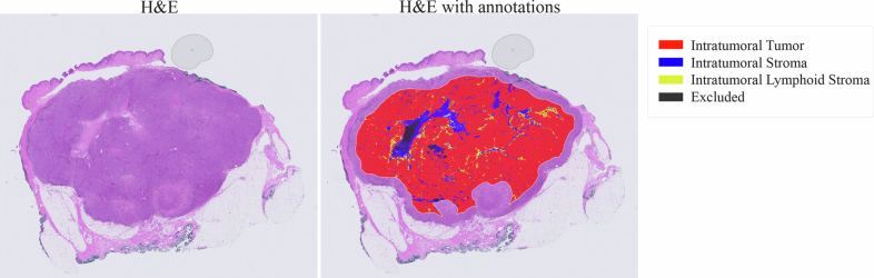
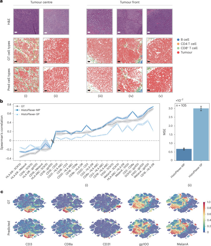
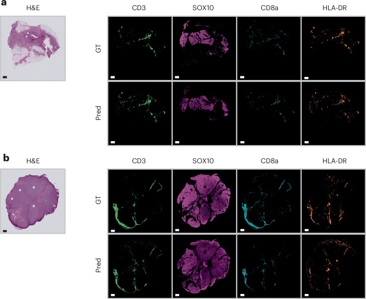
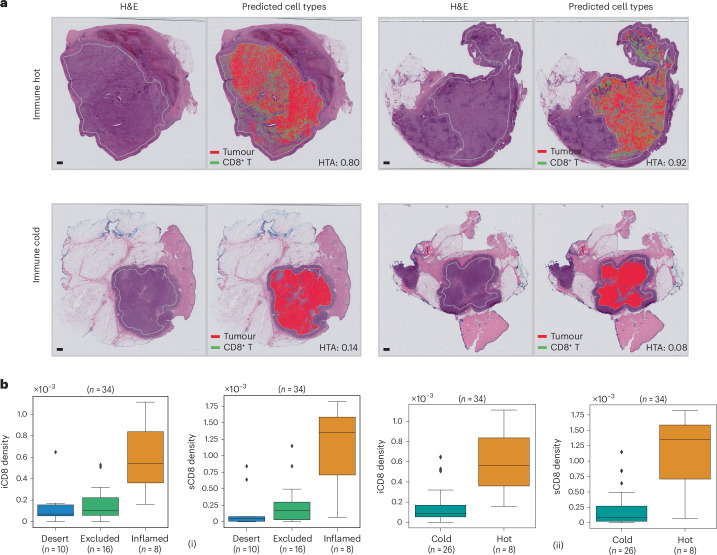
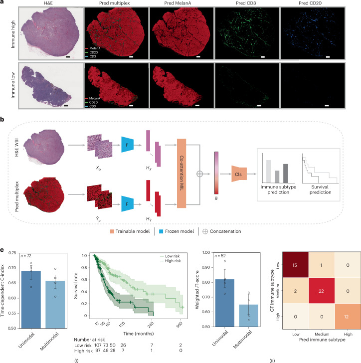
Multiplexed protein imaging offers valuable insights into interactions between tumours and their surrounding tumour microenvironment, but its widespread use is limited by cost, time and tissue availability. Here we present HistoPlexer, a deep learning framework that generates spatially resolved protein multiplexes directly from standard haematoxylin and eosin (H&E) histopathology images. HistoPlexer jointly predicts multiple tumour and immune markers using a conditional generative adversarial architecture with custom loss functions designed to ensure pixel- and embedding-level similarity while mitigating slice-to-slice variations. A comprehensive evaluation of metastatic melanoma samples demonstrates that HistoPlexer-generated protein maps closely resemble real maps, as validated by expert assessment. They preserve crucial biological relationships by capturing spatial co-localization patterns among proteins. The spatial distribution of immune infiltration from HistoPlexer-generated protein multiplex enables stratification of tumours into immune subtypes. In an independent cohort, integration of HistoPlexer-derived features into predictive models enhances performance in survival prediction and immune subtype classification compared to models using H&E features alone. To assess broader applicability, we benchmarked HistoPlexer on publicly available pixel-aligned datasets from different cancer types. In all settings, HistoPlexer consistently outperformed baseline methods, demonstrating robustness across diverse tissue types and imaging conditions. By enabling whole-slide protein multiplex generation from routine H&E images, HistoPlexer offers a cost- and time-efficient approach to tumour microenvironment characterization with strong potential to advance precision oncology.
多重蛋白质成像为了解肿瘤与其周围肿瘤微环境之间的相互作用提供了有价值的见解,但其广泛应用受到成本、时间和组织可用性的限制。在此,我们展示了HistoPlexer,这是一种深度学习框架,可直接从标准苏木精和伊红(H&E)组织病理学图像生成空间分辨的蛋白质多重图像。HistoPlexer使用条件生成对抗架构联合预测多种肿瘤和免疫标志物,并设计了自定义损失函数,以确保像素级和嵌入级的相似性,同时减轻切片间的差异。对转移性黑色素瘤样本的全面评估表明,经专家评估验证,HistoPlexer生成的蛋白质图谱与真实图谱非常相似。它们通过捕捉蛋白质之间的空间共定位模式,保留了关键的生物学关系。HistoPlexer生成的蛋白质多重图像中的免疫浸润空间分布能够将肿瘤分层为免疫亚型。在一个独立队列中,与仅使用H&E特征的模型相比,将HistoPlexer衍生的特征整合到预测模型中可提高生存预测和免疫亚型分类的性能。为了评估更广泛的适用性,我们在来自不同癌症类型的公开可用像素对齐数据集上对HistoPlexer进行了基准测试。在所有情况下,HistoPlexer始终优于基线方法,证明了其在不同组织类型和成像条件下的稳健性。通过从常规H&E图像中生成全切片蛋白质多重图像,HistoPlexer提供了一种经济高效的肿瘤微环境表征方法,具有推进精准肿瘤学的强大潜力。