Muñoz Manuel F, Matsumoto Collin, Rhana Paula, Collier Daniel M, Santana L Fernando
Department of Physiology & Membrane Biology, School of Medicine, University of California, Davis, USA.
bioRxiv. 2025 Aug 22:2025.08.18.670947. doi: 10.1101/2025.08.18.670947.
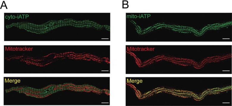
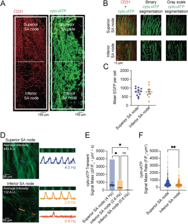
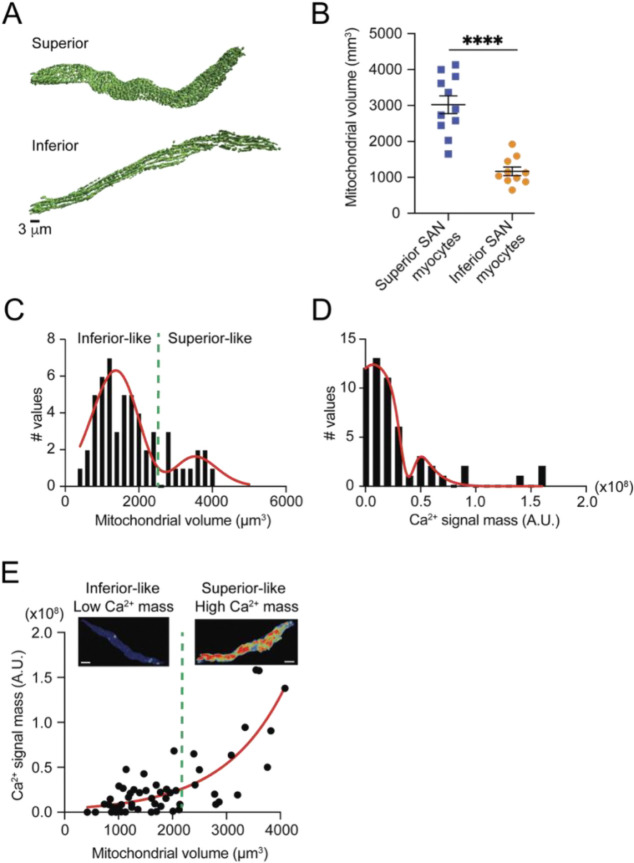
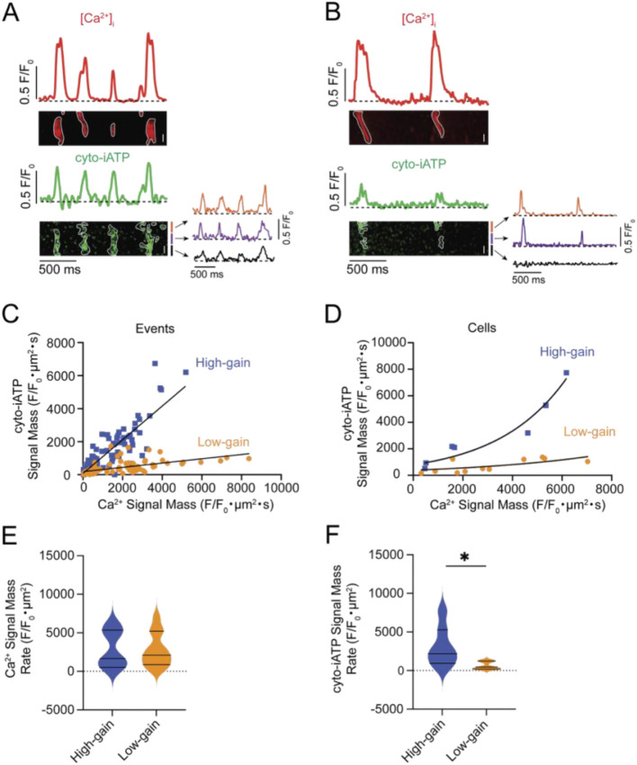
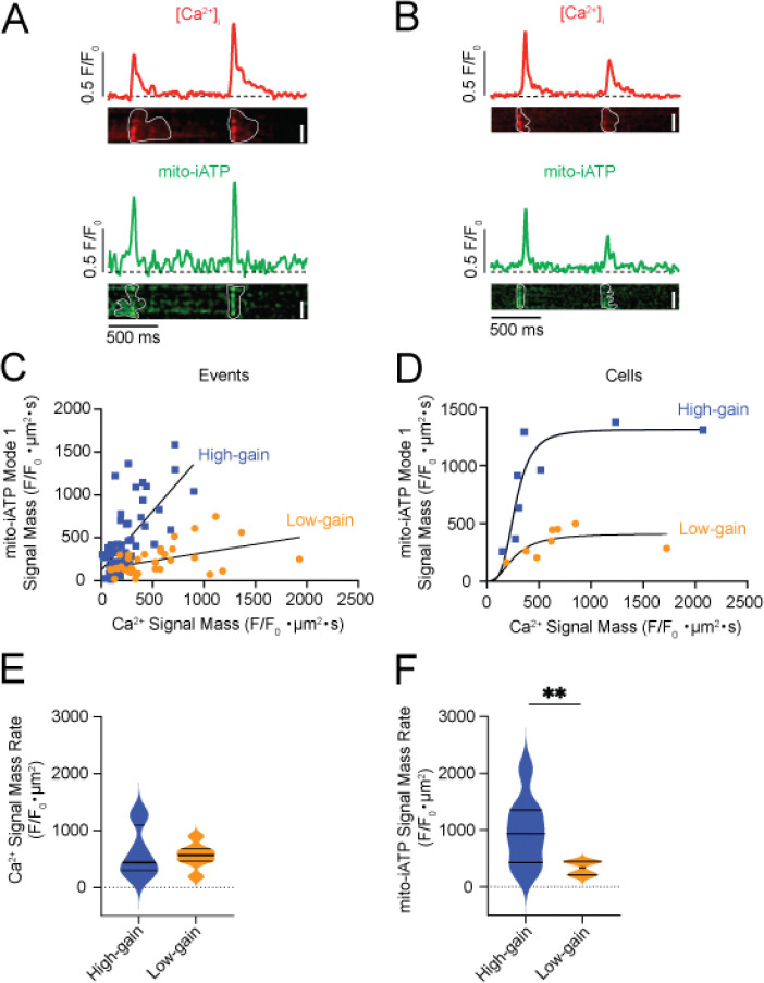
Pacemaker myocytes of the sinoatrial (SA) node initiate each heartbeat through coupled voltage and Ca oscillators, but whether ATP supply is regulated on a beat-by-beat schedule in these cells has been unclear. Using genetically encoded sensors targeted to the cytosol and mitochondria, we tracked beat-resolved ATP dynamics in intact mouse SA node and isolated myocytes. Cytosolic ATP rose transiently with each Ca transient and segregated into high- and low-gain phenotypes defined by the Ca-ATP coupling slope. Mitochondrial ATP flux adopted two stereotyped waveforms-Mode-1 "gains" and Mode-2 "dips." Within Mode-1 cells, ATP gains mirrored the cytosolic high/low-gain dichotomy; Mode-2 dips scaled linearly with Ca load and predominated in slower-firing cells. In the intact node, high-gain/Mode-1 phenotypes localized to superior regions and low-gain/Mode-2 to inferior regions, paralleling gradients in rate, mitochondrial volume, and capillary density. Pharmacology placed the Ca clock upstream of ATP production: the HCN channel blocker ivabradine slowed the ATP cycle without changing amplitude, whereas the SERCA pump inhibitor thapsigargin or the mitochondrial uncoupler FCCP abolished transients. Mode-2 recovery kinetics indicate slower ATP replenishment that would favor low-frequency, fluctuation-rich firing in a subset of cells. Together, these findings reveal beat-locked metabolic microdomains in which the Ca clock times oxidative phosphorylation under a local O ceiling, unifying vascular architecture, mitochondrial organization, and Ca signaling to coordinate energy supply with excitability. This energetic hierarchy helps explain why some SA node myocytes are more likely to set rate whereas others may widen bandwidth.
窦房结(SA)的起搏心肌细胞通过耦合电压和钙振荡器启动每次心跳,但这些细胞中的ATP供应是否在逐搏计划中受到调节尚不清楚。我们使用靶向细胞质和线粒体的基因编码传感器,追踪完整小鼠窦房结和分离心肌细胞中逐搏解析的ATP动态。细胞质ATP随着每次钙瞬变而短暂升高,并分为由钙-ATP耦合斜率定义的高增益和低增益表型。线粒体ATP通量采用两种定型波形——模式1“增益”和模式2“下降”。在模式1细胞中,ATP增益反映了细胞质高/低增益二分法;模式2下降与钙负荷呈线性比例,在放电较慢的细胞中占主导地位。在完整的节点中,高增益/模式1表型定位于上部区域,低增益/模式2定位于下部区域,与速率、线粒体体积和毛细血管密度的梯度平行。药理学研究表明钙时钟位于ATP产生的上游:HCN通道阻滞剂伊伐布雷定减慢了ATP循环而不改变幅度,而SERCA泵抑制剂毒胡萝卜素或线粒体解偶联剂FCCP消除了瞬变。模式2恢复动力学表明ATP补充较慢,这有利于一部分细胞进行低频、富含波动的放电。总之,这些发现揭示了节拍锁定的代谢微区,其中钙时钟在局部氧上限下调节氧化磷酸化,将血管结构、线粒体组织和钙信号统一起来,以协调能量供应与兴奋性。这种能量层次结构有助于解释为什么一些窦房结心肌细胞更有可能设定心率,而另一些细胞可能会拓宽带宽。